CT scan showing kidney cancer (axial section)
Bildnummer 11837798
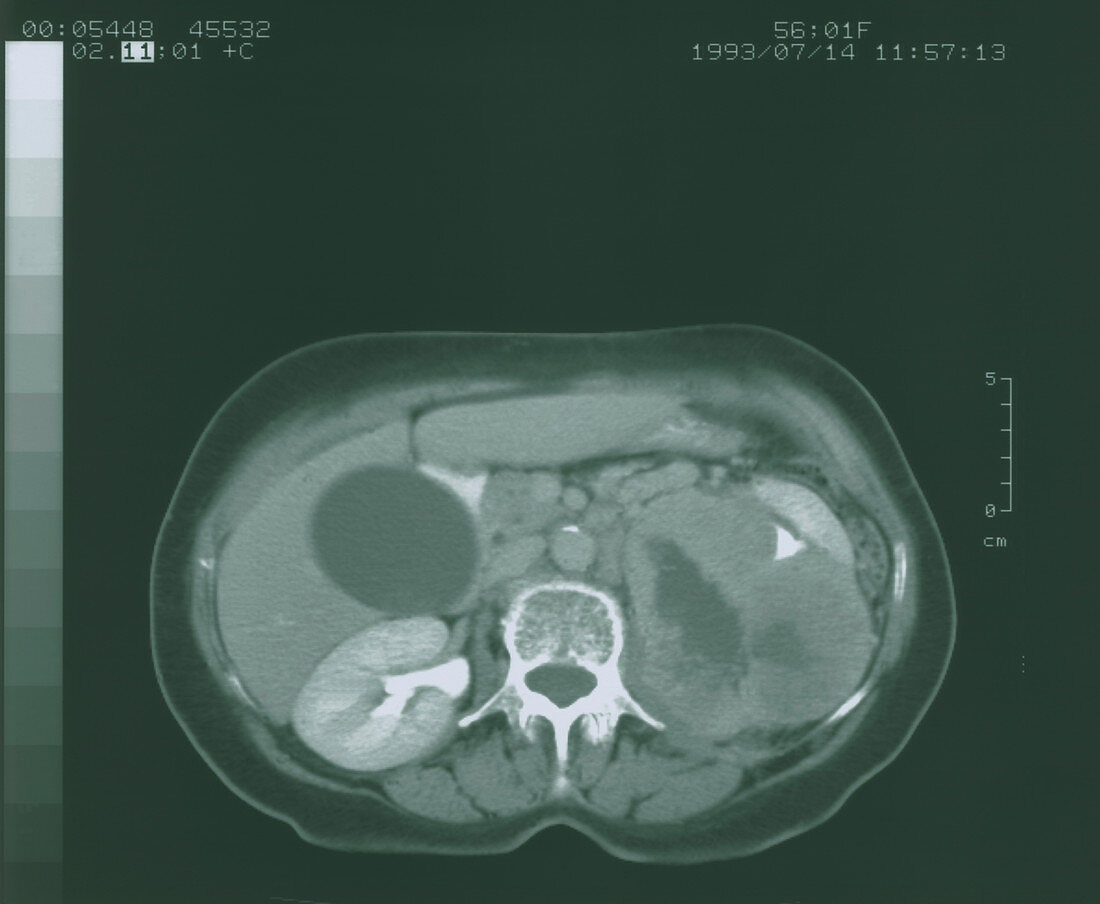
| Kidney cancer. Computed Tomography (CT) scan of an axial section through the human abdomen,showing kidney cancer. At centre is a vertebral bone of the spin (white). The diseased kidney is distorted and enlarged by a cancer tumour (at right). This diseased kidney with dark coloured interior is largely non-functioning; it has a small function- ing area excreting into the collecting system (white triangle,upper right). At lower left is a normal kidney,bean-shaped,that is excreting an X-ray contrast agent (white). The liver (extreme left) is seen next to the normal kidney,with the dark circular gall bladder covering the liver | |
| Lizenzart: | Lizenzpflichtig |
| Credit: | Science Photo Library |
| Bildgröße: | 4606 px × 3785 px |
| Modell-Rechte: | nicht erforderlich |
| Eigentums-Rechte: | nicht erforderlich |
| Restrictions: | - |
Preise für dieses Bild ab 15 €
Universitäten & Organisationen
(Informationsmaterial Digital, Informationsmaterial Print, Lehrmaterial Digital etc.)
ab 15 €
Redaktionell
(Bücher, Bücher: Sach- und Fachliteratur, Digitale Medien (redaktionell) etc.)
ab 30 €
Werbung
(Anzeigen, Aussenwerbung, Digitale Medien, Fernsehwerbung, Karten, Werbemittel, Zeitschriften etc.)
ab 55 €
Handelsprodukte
(bedruckte Textilie, Kalender, Postkarte, Grußkarte, Verpackung etc.)
ab 75 €
Pauschalpreise
Rechtepakete für die unbeschränkte Bildnutzung in Print oder Online
ab 495 €