CT scan showing a large brain tumour
Bildnummer 11837795
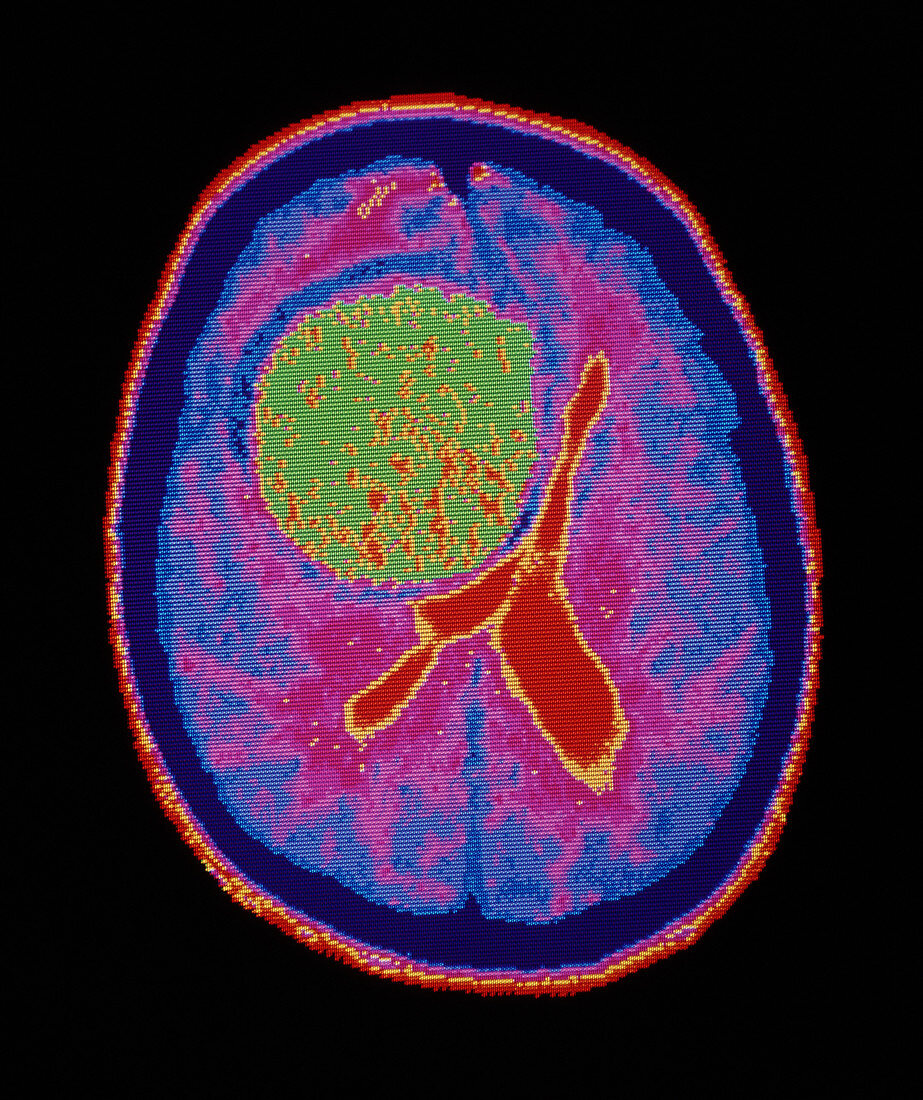
| Coloured computed tomography (CT) brain scan showing a large,intracranial tumour,the large round mass (green) seen at centre. Within the tumour,spots indicate fluid-filled cysts. The outer border of the tumour (pink) has become calcified with age. This tumour is most probably a low-grade glioma,a tumour of the supporting glial cells,the specialised connective tissue of the brain and central nervous system. Gliomas include the astrocytomas and glioblastomas and account for the majority of intracranial tumours. Slow-growing,low-grade (less malignant) gliomas may exist for years before rapidly acquiring a malignant character | |
| Lizenzart: | Lizenzpflichtig |
| Credit: | Science Photo Library / Kulyk, Mehau |
| Bildgröße: | 3453 px × 4115 px |
| Modell-Rechte: | nicht erforderlich |
| Eigentums-Rechte: | nicht erforderlich |
| Restrictions: | - |
Preise für dieses Bild ab 15 €
Universitäten & Organisationen
(Informationsmaterial Digital, Informationsmaterial Print, Lehrmaterial Digital etc.)
ab 15 €
Redaktionell
(Bücher, Bücher: Sach- und Fachliteratur, Digitale Medien (redaktionell) etc.)
ab 30 €
Werbung
(Anzeigen, Aussenwerbung, Digitale Medien, Fernsehwerbung, Karten, Werbemittel, Zeitschriften etc.)
ab 55 €
Handelsprodukte
(bedruckte Textilie, Kalender, Postkarte, Grußkarte, Verpackung etc.)
ab 75 €
Pauschalpreise
Rechtepakete für die unbeschränkte Bildnutzung in Print oder Online
ab 495 €