Secondary cancer,bone scan
Bildnummer 11837769
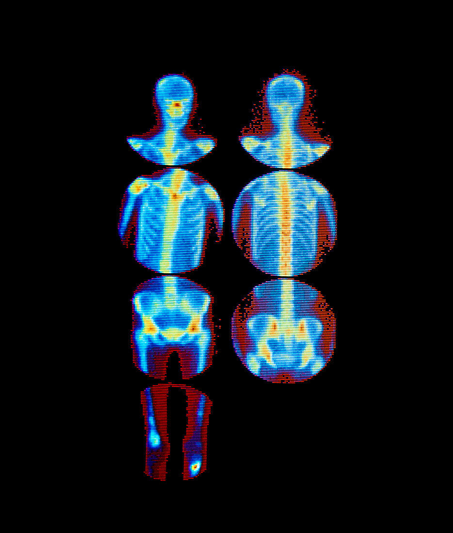
| Secondary (metastatic) bone cancer: false-colour gamma camera scans of the human skeleton in anterior (left) and posterior views. In the anterior view,the image of the lower legs (bottom left) shows an increased uptake of radioactivity in the region of the right femur (left on image) and the left tibia. This is consistent with the sites of bone metastases,given that the patient had already received treatment for a primary cancer. This diagnosis would be confirmed by X-ray examination. The radioactive tracer used here was technetium-99m linked to methylene diphosphate (MDP) | |
| Lizenzart: | Lizenzpflichtig |
| Credit: | Science Photo Library / MEDICAL PHYSICS, RVI, NEWCASTLE UPON-TYNE / SIMON FRASER |
| Bildgröße: | 4212 px × 4961 px |
| Modell-Rechte: | nicht erforderlich |
| Eigentums-Rechte: | nicht erforderlich |
| Restrictions: | - |
Preise für dieses Bild ab 15 €
Universitäten & Organisationen
(Informationsmaterial Digital, Informationsmaterial Print, Lehrmaterial Digital etc.)
ab 15 €
Redaktionell
(Bücher, Bücher: Sach- und Fachliteratur, Digitale Medien (redaktionell) etc.)
ab 30 €
Werbung
(Anzeigen, Aussenwerbung, Digitale Medien, Fernsehwerbung, Karten, Werbemittel, Zeitschriften etc.)
ab 55 €
Handelsprodukte
(bedruckte Textilie, Kalender, Postkarte, Grußkarte, Verpackung etc.)
ab 75 €
Pauschalpreise
Rechtepakete für die unbeschränkte Bildnutzung in Print oder Online
ab 495 €