Cancer cell
Bildnummer 11837468
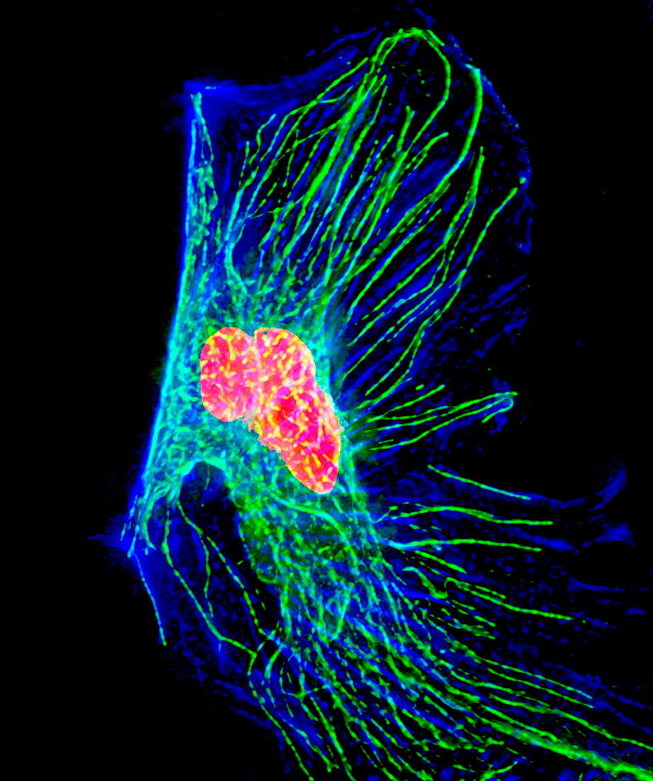
| Cancer cell. Immunofluorescent light micrograph of a cancer cell. The abnormal distribution of DNA (yellow/red),microtubules (green) and actin (blue) is shown. Immunofluorescence uses antibodies to attach fluorescent dyes to specific cell tissues | |
| Lizenzart: | Lizenzpflichtig |
| Credit: | Science Photo Library / DR PAUL ANDREWS, UNIVERSITY OF DUNDEE |
| Bildgröße: | 2304 px × 2754 px |
| Modell-Rechte: | nicht erforderlich |
| Eigentums-Rechte: | nicht erforderlich |
| Restrictions: | - |
Preise für dieses Bild ab 15 €
Universitäten & Organisationen
(Informationsmaterial Digital, Informationsmaterial Print, Lehrmaterial Digital etc.)
ab 15 €
Redaktionell
(Bücher, Bücher: Sach- und Fachliteratur, Digitale Medien (redaktionell) etc.)
ab 30 €
Werbung
(Anzeigen, Aussenwerbung, Digitale Medien, Fernsehwerbung, Karten, Werbemittel, Zeitschriften etc.)
ab 55 €
Handelsprodukte
(bedruckte Textilie, Kalender, Postkarte, Grußkarte, Verpackung etc.)
ab 75 €
Pauschalpreise
Rechtepakete für die unbeschränkte Bildnutzung in Print oder Online
ab 495 €