Immunofluorescent LM of a melanoma cancer cell
Bildnummer 11837170
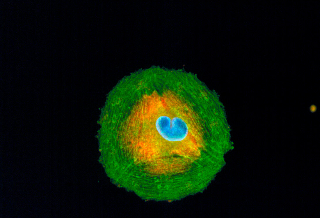
| Melanoma cell. Immunofluorescent Light Micrograph of a melanoma cancer cell,cultured from a human tumour. The cell nucleus (blue) is in the process of division; cytoplasm stains orange; actin stress fibres (green,protein) surround the cell & serve as a supportive network. This cancer is typically derived from melanin-forming skin cells. Melanoma is a highly malignant cancer consisting of large undifferentiated cells that divide rapidly,and invade healthy tissue. Immunofluorescence is a technique which uses antibodies to attach fluores- cent dyes to specific tissues and molecules within the cell. Magnification: x125 at 35mm size | |
| Lizenzart: | Lizenzpflichtig |
| Credit: | Science Photo Library / Kedersha, Nancy |
| Bildgröße: | 3700 px × 2516 px |
| Modell-Rechte: | nicht erforderlich |
| Eigentums-Rechte: | nicht erforderlich |
| Restrictions: | - |
Preise für dieses Bild ab 15 €
Universitäten & Organisationen
(Informationsmaterial Digital, Informationsmaterial Print, Lehrmaterial Digital etc.)
ab 15 €
Redaktionell
(Bücher, Bücher: Sach- und Fachliteratur, Digitale Medien (redaktionell) etc.)
ab 30 €
Werbung
(Anzeigen, Aussenwerbung, Digitale Medien, Fernsehwerbung, Karten, Werbemittel, Zeitschriften etc.)
ab 55 €
Handelsprodukte
(bedruckte Textilie, Kalender, Postkarte, Grußkarte, Verpackung etc.)
ab 75 €
Pauschalpreise
Rechtepakete für die unbeschränkte Bildnutzung in Print oder Online
ab 495 €