Immunofluorescent LM of mixed human-cancer cells
Bildnummer 11837158
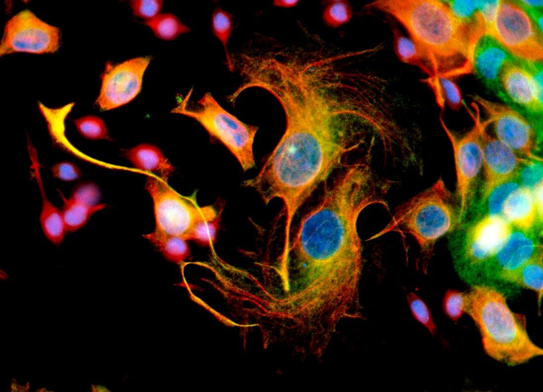
| Immunofluorescent Light Micrograph of mixed human- cancer cells in a culture: squamous carcinoma (at left),leukaemia (centre),& epithelial carcinoma (right). Cell nuclei stain blue. These cancers can be highly malignant,rapidly dividing growth forms. Both squamous (green) and epithelial (red) carcinoma occur on body surfaces: skin,or cells lining internal body organs. While leukaemia (orange) is a cancer of blood and blood-forming organs. Immunofluorescence is a staining technique which uses antibodies to attach fluorescent dyes to specific tissues and to molecules in the cell. Magnification: x400 at 35mm,x675 at 6x4.5cm size | |
| Lizenzart: | Lizenzpflichtig |
| Credit: | Science Photo Library / Kedersha, Nancy |
| Bildgröße: | 4935 px × 3564 px |
| Modell-Rechte: | nicht erforderlich |
| Eigentums-Rechte: | nicht erforderlich |
| Restrictions: | - |
Preise für dieses Bild ab 15 €
Universitäten & Organisationen
(Informationsmaterial Digital, Informationsmaterial Print, Lehrmaterial Digital etc.)
ab 15 €
Redaktionell
(Bücher, Bücher: Sach- und Fachliteratur, Digitale Medien (redaktionell) etc.)
ab 30 €
Werbung
(Anzeigen, Aussenwerbung, Digitale Medien, Fernsehwerbung, Karten, Werbemittel, Zeitschriften etc.)
ab 55 €
Handelsprodukte
(bedruckte Textilie, Kalender, Postkarte, Grußkarte, Verpackung etc.)
ab 75 €
Pauschalpreise
Rechtepakete für die unbeschränkte Bildnutzung in Print oder Online
ab 495 €