LM of melanoma cancer cells
Bildnummer 11837154
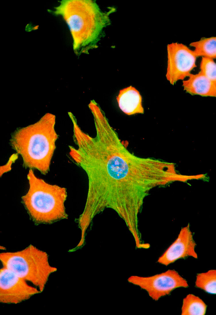
| Immunofluorescent Light Micrograph of melanoma cancer cells and fibroblasts,cultured from a human tumour. A fibroblast (star-shaped cell,at centre),forms connective tissue to support other cells. Actin fibres (green) around it help provide this supportive network. Smaller melanoma cells have a large nucleus (blue); cytoplasm is orange. Derived from melanin-forming cells of the skin,they are highly malignant & divide rapidly (seen at upper right). Immunofluorescence is a staining technique which uses antibodies to attach fluorescent dyes to specific tissues and to molecules within the cell.Mag:x200 at35mm,x340 at 6x4.5size | |
| Lizenzart: | Lizenzpflichtig |
| Credit: | Science Photo Library / Kedersha, Nancy |
| Bildgröße: | 2488 px × 3633 px |
| Modell-Rechte: | nicht erforderlich |
| Eigentums-Rechte: | nicht erforderlich |
| Restrictions: | - |
Preise für dieses Bild ab 15 €
Universitäten & Organisationen
(Informationsmaterial Digital, Informationsmaterial Print, Lehrmaterial Digital etc.)
ab 15 €
Redaktionell
(Bücher, Bücher: Sach- und Fachliteratur, Digitale Medien (redaktionell) etc.)
ab 30 €
Werbung
(Anzeigen, Aussenwerbung, Digitale Medien, Fernsehwerbung, Karten, Werbemittel, Zeitschriften etc.)
ab 55 €
Handelsprodukte
(bedruckte Textilie, Kalender, Postkarte, Grußkarte, Verpackung etc.)
ab 75 €
Pauschalpreise
Rechtepakete für die unbeschränkte Bildnutzung in Print oder Online
ab 495 €