Arachnoid cyst,MRI scan
Bildnummer 11836533
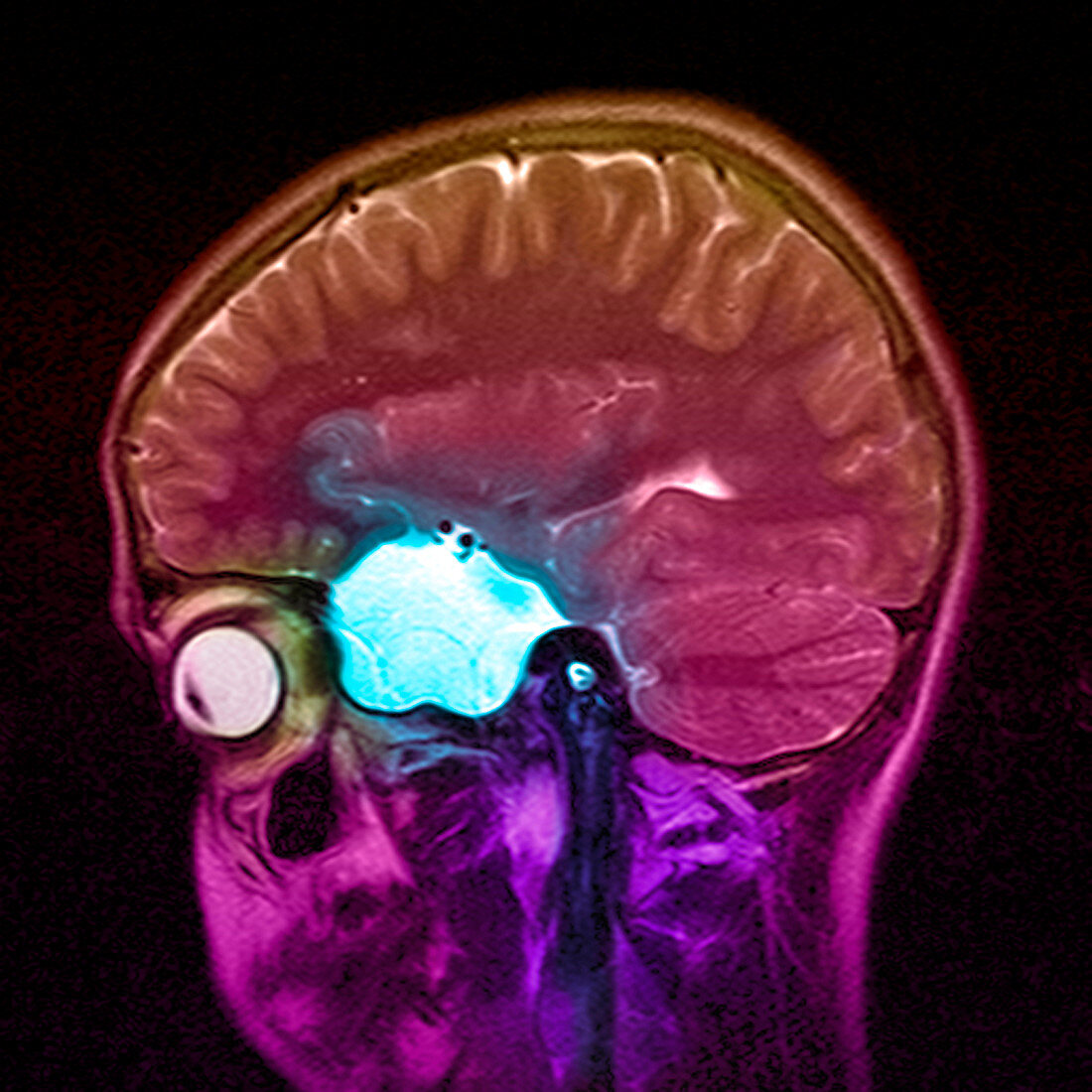
| Arachnoid cyst. Coloured magnetic resonance imaging (MRI) scan of a sagittal (side) section through the head of a 13 year old boy. The front of the head is at left. The scan shows a large left middle cranial fossa arachnoid cyst (blue). This is an abnormal collection of CSF (cerebral spinal fluid),which is walled off and acts like a mass. It is benign but can cause a variety of symptoms including seizures. MRI uses radio waves and a magnet to obtain "slice" body images | |
| Lizenzart: | Lizenzpflichtig |
| Credit: | Science Photo Library / Fraser, Simon |
| Bildgröße: | 3543 px × 3543 px |
| Modell-Rechte: | nicht erforderlich |
| Eigentums-Rechte: | nicht erforderlich |
| Restrictions: | - |
Preise für dieses Bild ab 15 €
Universitäten & Organisationen
(Informationsmaterial Digital, Informationsmaterial Print, Lehrmaterial Digital etc.)
ab 15 €
Redaktionell
(Bücher, Bücher: Sach- und Fachliteratur, Digitale Medien (redaktionell) etc.)
ab 30 €
Werbung
(Anzeigen, Aussenwerbung, Digitale Medien, Fernsehwerbung, Karten, Werbemittel, Zeitschriften etc.)
ab 55 €
Handelsprodukte
(bedruckte Textilie, Kalender, Postkarte, Grußkarte, Verpackung etc.)
ab 75 €
Pauschalpreise
Rechtepakete für die unbeschränkte Bildnutzung in Print oder Online
ab 495 €
Keywords
- Arachnoidalzyste,
- csf,
- Diagnose,
- diagnostizieren,
- eingefärbt,
- Epilepsie,
- farbig,
- Fossa,
- gefärbt,
- Gehirn,
- gutartig,
- Hirnscan,
- Jugend,
- Jugendlicher,
- Junge,
- Kind,
- Kondition,
- Krankheit,
- Magnetresonanzbildgebung,
- Magnetresonanztomografie,
- Männlich,
- Medizin,
- medizinisch,
- medizinisches Bild,
- Mensch,
- Menschen,
- menschlicher Körper,
- MRI,
- MRT-Untersuchung,
- Neuroimaging,
- Neurologie,
- neurologisch,
- Organ,
- Person,
- Querschnitt,
- Scanner,
- Schädel,
- Seite,
- Sektion,
- sektioniert,
- Störung,
- Teenager,
- Teenageralter,
- Temporallappen,
- Wissenschaft,
- Zyste