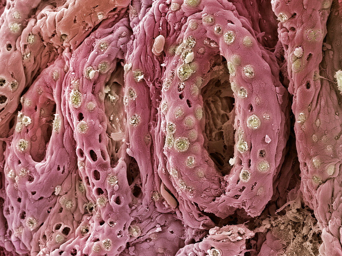
Ulcerative colitis,SEM
Bildnummer 11836299

| Ulcerative colitis. Coloured scanning electron micrograph (SEM) of goblet cells (green) on the mucosal surface of the bowel of a patient suffering from ulcerative colitis. The intestinal wall contains the tubular glands (pink) seen here. An orifice of one of these glands is at centre left. The irregular appearance of these structures may be due to ulcerative colitis. The pink surface is covered with numerous microvilli (not seen) that absorb nutrients and secrete digestive substances. The goblet cells secrete the mucus that lines the intestinal tract. Ulcerative colitis is the inflammation and ulceration of the colon and rectum. Magnification unknown | |
| Lizenzart: | Lizenzpflichtig |
| Credit: | Science Photo Library / Gschmeissner, Steve |
| Bildgröße: | 2560 px × 1920 px |
| Modell-Rechte: | nicht erforderlich |
| Eigentums-Rechte: | nicht erforderlich |
| Restrictions: | - |
Preise für dieses Bild ab 15 €
Universitäten & Organisationen
(Informationsmaterial Digital, Informationsmaterial Print, Lehrmaterial Digital etc.)
ab 15 €
Redaktionell
(Bücher, Bücher: Sach- und Fachliteratur, Digitale Medien (redaktionell) etc.)
ab 30 €
Werbung
(Anzeigen, Aussenwerbung, Digitale Medien, Fernsehwerbung, Karten, Werbemittel, Zeitschriften etc.)
ab 55 €
Handelsprodukte
(bedruckte Textilie, Kalender, Postkarte, Grußkarte, Verpackung etc.)
ab 75 €
Pauschalpreise
Rechtepakete für die unbeschränkte Bildnutzung in Print oder Online
ab 495 €
