MRI scan of human brain diseased with CJD
Bildnummer 11836194
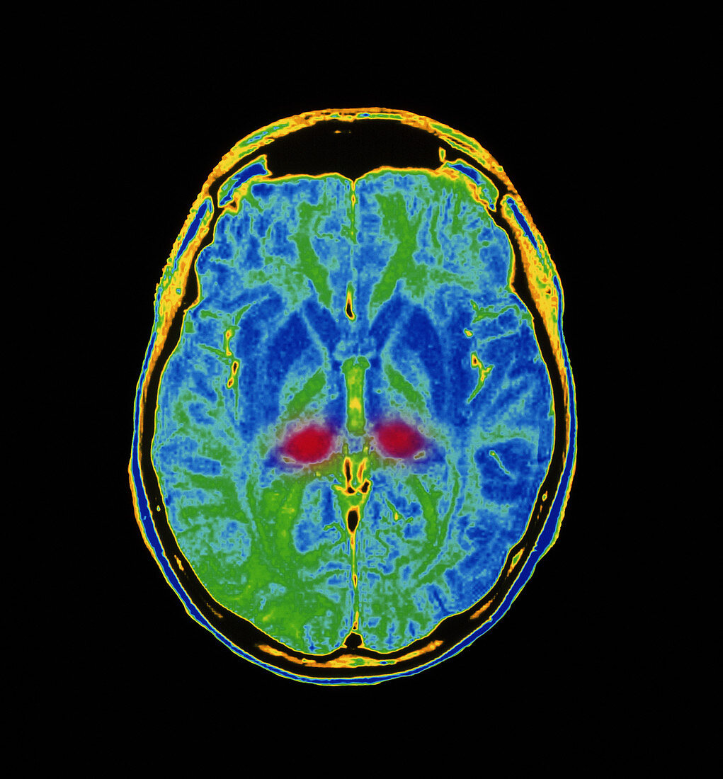
| Creutzfeldt-Jakob disease. Coloured Magnetic Resonance Imaging (MRI) scan of the brain of a 17 year old male suffering from Creutzfeldt-Jakob disease (CJD: new variant) in 1997. The patient died. The front of the head is at top. In this axial "slice" through the brain,the folded cerebrum is seen forming two hemispheres. At lower centre are two red areas of the thalamus diseased with CJD. It is detected as a 'bilateral signal abnormality' on MRI. CJD destroys nerve cells and causes brain tissue to become spongy. Symptoms include dementia and muscle contractions,and death. There is concern that eating beef from "mad cows" may cause CJD | |
| Lizenzart: | Lizenzpflichtig |
| Credit: | Science Photo Library / Fraser, Simon / Newcastle Upon Tyne / Royal Victoria Infirmary |
| Bildgröße: | 3630 px × 3912 px |
| Modell-Rechte: | nicht erforderlich |
| Eigentums-Rechte: | nicht erforderlich |
| Restrictions: | - |
Preise für dieses Bild ab 15 €
Universitäten & Organisationen
(Informationsmaterial Digital, Informationsmaterial Print, Lehrmaterial Digital etc.)
ab 15 €
Redaktionell
(Bücher, Bücher: Sach- und Fachliteratur, Digitale Medien (redaktionell) etc.)
ab 30 €
Werbung
(Anzeigen, Aussenwerbung, Digitale Medien, Fernsehwerbung, Karten, Werbemittel, Zeitschriften etc.)
ab 55 €
Handelsprodukte
(bedruckte Textilie, Kalender, Postkarte, Grußkarte, Verpackung etc.)
ab 75 €
Pauschalpreise
Rechtepakete für die unbeschränkte Bildnutzung in Print oder Online
ab 495 €