Coloured SEM of a liver cell affected by cirrhosis
Bildnummer 11836126
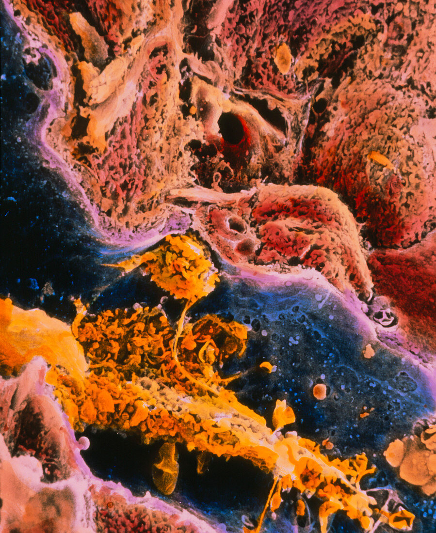
| Cirrhosis. Coloured scanning electron micrograph of a human liver cell (red-brown),known as hepatocyte,showing abnormal features caused by cirrhosis. It is a disease in which bands of connective tissue (pale brown upper centre and top left) break up the structure of the liver severely impairing its functions. The surface of the liver cell (upper centre right and left) is abnormal showing irregular microvilli and larger lacunae. Two activated macrophages (Kupffer cells yellow) are seen in a capillary (blue). The wall of the capillary (pale purple) is very thick and fenestrated. Magnification: x1650 at 6x7cm size | |
| Lizenzart: | Lizenzpflichtig |
| Credit: | Science Photo Library / PROFESSORS P.M. MOTTA & T. FUJITA |
| Bildgröße: | 2948 px × 3609 px |
| Modell-Rechte: | nicht erforderlich |
| Eigentums-Rechte: | nicht erforderlich |
| Restrictions: | - |
Preise für dieses Bild ab 15 €
Universitäten & Organisationen
(Informationsmaterial Digital, Informationsmaterial Print, Lehrmaterial Digital etc.)
ab 15 €
Redaktionell
(Bücher, Bücher: Sach- und Fachliteratur, Digitale Medien (redaktionell) etc.)
ab 30 €
Werbung
(Anzeigen, Aussenwerbung, Digitale Medien, Fernsehwerbung, Karten, Werbemittel, Zeitschriften etc.)
ab 55 €
Handelsprodukte
(bedruckte Textilie, Kalender, Postkarte, Grußkarte, Verpackung etc.)
ab 75 €
Pauschalpreise
Rechtepakete für die unbeschränkte Bildnutzung in Print oder Online
ab 495 €