Artwork showing rheumatoid arthritis of the knee
Bildnummer 11835130
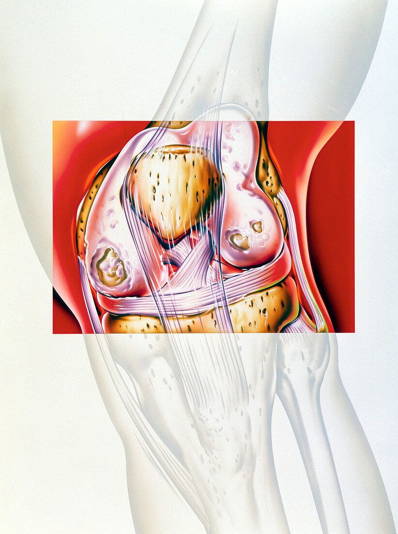
| Arthritis. Artwork of a front view of a human knee joint affected by rheumatoid arthritis. The knee cap (patella,yellow) is at upper centre,between the thigh bone (femur,above),and the shin bone (tibia) and fibula (below). The protective layer of cartilage (pink) covering the joint has worn away in patches. The fibrous pink tissues are tendons and ligaments,which hold muscles and bones together inside joints. Rheumatoid arthritis occurs when the immune system attacks the bones and soft tissues of a joint. The joints may become swollen,painful,stiff and deformed. Treatment is with anti-inflammatory drugs | |
| Lizenzart: | Lizenzpflichtig |
| Credit: | Science Photo Library / Bavosi, John |
| Bildgröße: | 2644 px × 3543 px |
| Modell-Rechte: | nicht erforderlich |
| Eigentums-Rechte: | nicht erforderlich |
| Restrictions: | - |
Preise für dieses Bild ab 15 €
Universitäten & Organisationen
(Informationsmaterial Digital, Informationsmaterial Print, Lehrmaterial Digital etc.)
ab 15 €
Redaktionell
(Bücher, Bücher: Sach- und Fachliteratur, Digitale Medien (redaktionell) etc.)
ab 30 €
Werbung
(Anzeigen, Aussenwerbung, Digitale Medien, Fernsehwerbung, Karten, Werbemittel, Zeitschriften etc.)
ab 55 €
Handelsprodukte
(bedruckte Textilie, Kalender, Postkarte, Grußkarte, Verpackung etc.)
ab 75 €
Pauschalpreise
Rechtepakete für die unbeschränkte Bildnutzung in Print oder Online
ab 495 €