Osteoarthritic knee
Bildnummer 11835093
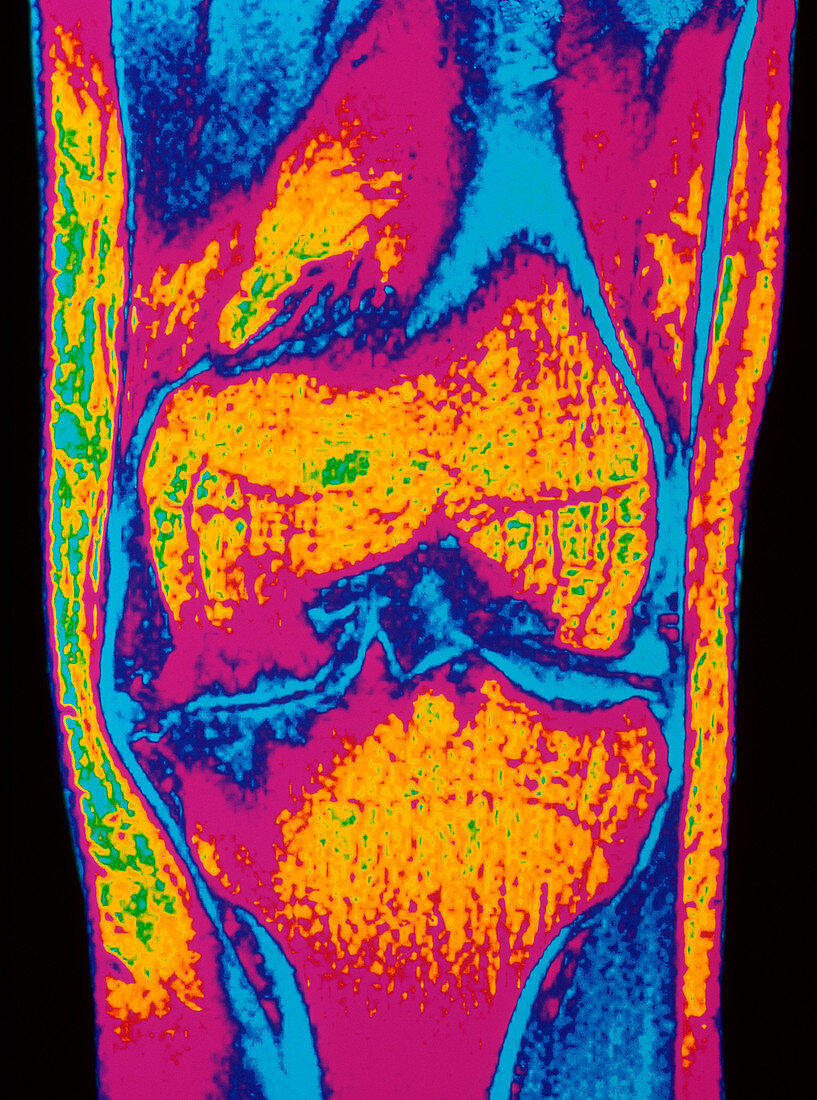
| Osteoarthritis of knee. Colour Magnetic Resonance Imaging (MRI) scan of a front view of the knee joint of a 33 year old male with osteoarthritis. The lower end of the femur (thigh bone) is at centre (yellow); it articulates with the upper end of the tibia (shin bone) at lower centre (yellow). Due to osteoarthritis the ends of the bones have become severely eroded and jagged; cracking of the bone is also visible. A deep intercondylar notch (triangular) has formed in the femur. Osteo- arthritis is the most common type of arthritis due mainly to wear and tear on joints in the elderly. This patient had haemophilia which was a factor | |
| Lizenzart: | Lizenzpflichtig |
| Credit: | Science Photo Library / SIMON FRASER, ROYAL VICTORIA INFIRMARY |
| Bildgröße: | 3209 px × 4318 px |
| Modell-Rechte: | nicht erforderlich |
| Eigentums-Rechte: | nicht erforderlich |
| Restrictions: | - |
Preise für dieses Bild ab 15 €
Universitäten & Organisationen
(Informationsmaterial Digital, Informationsmaterial Print, Lehrmaterial Digital etc.)
ab 15 €
Redaktionell
(Bücher, Bücher: Sach- und Fachliteratur, Digitale Medien (redaktionell) etc.)
ab 30 €
Werbung
(Anzeigen, Aussenwerbung, Digitale Medien, Fernsehwerbung, Karten, Werbemittel, Zeitschriften etc.)
ab 55 €
Handelsprodukte
(bedruckte Textilie, Kalender, Postkarte, Grußkarte, Verpackung etc.)
ab 75 €
Pauschalpreise
Rechtepakete für die unbeschränkte Bildnutzung in Print oder Online
ab 495 €