Asthma pathology and treatment,diagram
Bildnummer 11834972
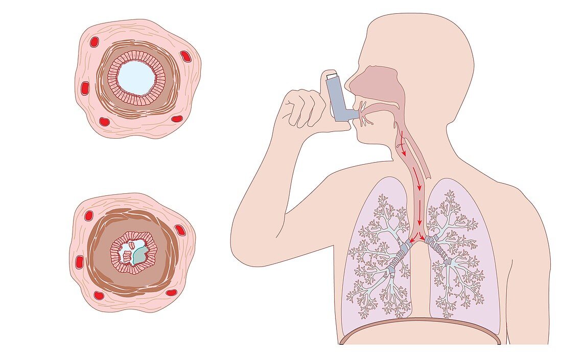
| Asthma pathology and treatment,cutaway diagrams. At right is an asthmatic patient using an inhaler to apply bronchodilator drugs to treat his asthma. The drugs (red arrows) are breathed in through the mouth,down the trachea and into the two lungs. The diagrams at left show cross-sections through a normal airway (top) and an asthmatic,constricted airway (bottom). The airways have a ring of smooth muscle (brown) and an inner layer of endothelial tissue (red/white). In the asthmatic airway,the smooth muscle has contracted,and the tissue is inflamed. Endothelial cell debris and mucus (blue) are blocking the airway. The bronchodilator drugs widen the airway,making it easier to breathe | |
| Lizenzart: | Lizenzpflichtig |
| Credit: | Science Photo Library / Gardiner, Peter |
| Bildgröße: | 3713 px × 2362 px |
| Modell-Rechte: | nicht erforderlich |
| Eigentums-Rechte: | nicht erforderlich |
| Restrictions: | - |
Preise für dieses Bild ab 15 €
Universitäten & Organisationen
(Informationsmaterial Digital, Informationsmaterial Print, Lehrmaterial Digital etc.)
ab 15 €
Redaktionell
(Bücher, Bücher: Sach- und Fachliteratur, Digitale Medien (redaktionell) etc.)
ab 30 €
Werbung
(Anzeigen, Aussenwerbung, Digitale Medien, Fernsehwerbung, Karten, Werbemittel, Zeitschriften etc.)
ab 55 €
Handelsprodukte
(bedruckte Textilie, Kalender, Postkarte, Grußkarte, Verpackung etc.)
ab 75 €
Pauschalpreise
Rechtepakete für die unbeschränkte Bildnutzung in Print oder Online
ab 495 €
Keywords
- Abschnitte,
- Alveolen,
- Anatomie,
- anatomisch,
- Arzneimittel,
- Asthma,
- Asthmaanfall,
- Asthmatiker,
- Atemweg,
- Atemwege,
- Atmungssystem,
- Ausrüstung,
- Behandlung,
- Bronchiole,
- Bronchiolen,
- Bronchodilatator,
- Cutaway,
- diagonal,
- Diagramm,
- Drogen,
- endothelial,
- geduldig,
- Gesundheitswesen,
- Illustration,
- Inhalator,
- Kondition,
- Kontraktion,
- Kunstwerk,
- Kunstwerke,
- Lumen,
- Lunge,
- Lungen,
- Medizin,
- medizinisch,
- Mensch,
- Menschen,
- menschlicher Körper,
- Pathologie,
- pathologisch,
- Person,
- Querschnitt,
- Querschnitte,
- Schicht,
- Schichten,
- Schleim,
- Sektion,
- sektioniert,
- Selbstbehandlung,
- Störung,
- Trümmer,
- verstopft,
- vorher,
- Zelle,
- Zellen