Artwork of inflamed bronchial epithelium in asthma
Bildnummer 11834772
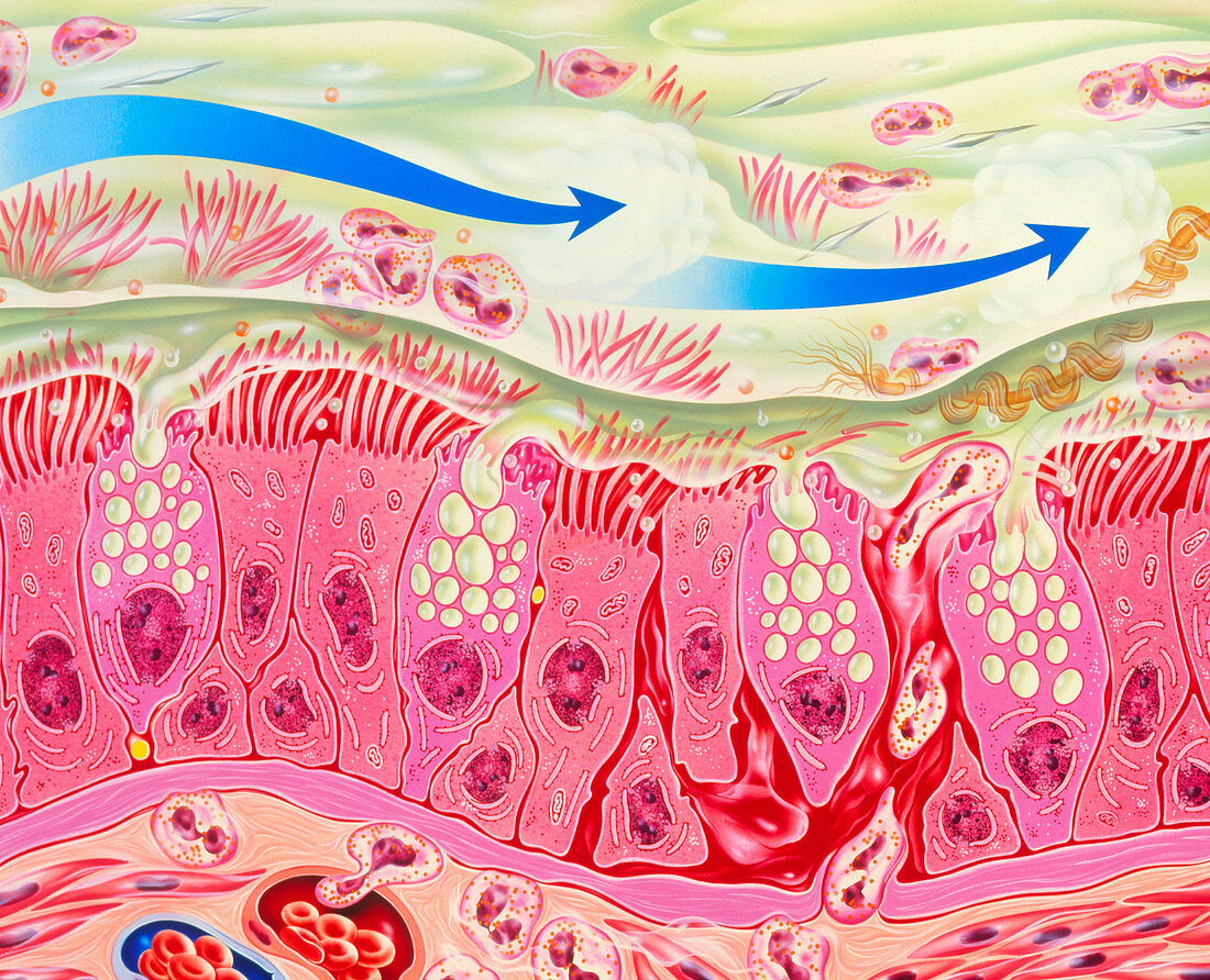
| Bronchial inflammation in asthma. Artwork of bron- chial epithelial tissue inflamed by asthma. A thick layer of mucus (green) covers the tissue. This is produced by goblet cells interspersed among ciliated epithelial cells. The connective tissue at the bottom contains enlarged blood vess- els (lower left) and numerous eosinophil white blood cells,which are also abundant in the mucus. Eosinophils play a major role in allergic inflam- mations. They secrete chemicals that are partly responsible for the bronchial constriction that occurs in asthma. Epithelial desquamation (shed- ding of outer layer of cells) is shown at right. The blue arrows symbolise drugs taken by inhaler | |
| Lizenzart: | Lizenzpflichtig |
| Credit: | Science Photo Library / Bavosi, John |
| Bildgröße: | 3424 px × 2778 px |
| Modell-Rechte: | nicht erforderlich |
| Eigentums-Rechte: | nicht erforderlich |
| Restrictions: | - |
Preise für dieses Bild ab 15 €
Universitäten & Organisationen
(Informationsmaterial Digital, Informationsmaterial Print, Lehrmaterial Digital etc.)
ab 15 €
Redaktionell
(Bücher, Bücher: Sach- und Fachliteratur, Digitale Medien (redaktionell) etc.)
ab 30 €
Werbung
(Anzeigen, Aussenwerbung, Digitale Medien, Fernsehwerbung, Karten, Werbemittel, Zeitschriften etc.)
ab 55 €
Handelsprodukte
(bedruckte Textilie, Kalender, Postkarte, Grußkarte, Verpackung etc.)
ab 75 €
Pauschalpreise
Rechtepakete für die unbeschränkte Bildnutzung in Print oder Online
ab 495 €