Sindbis virus,computer model
Bildnummer 11833743
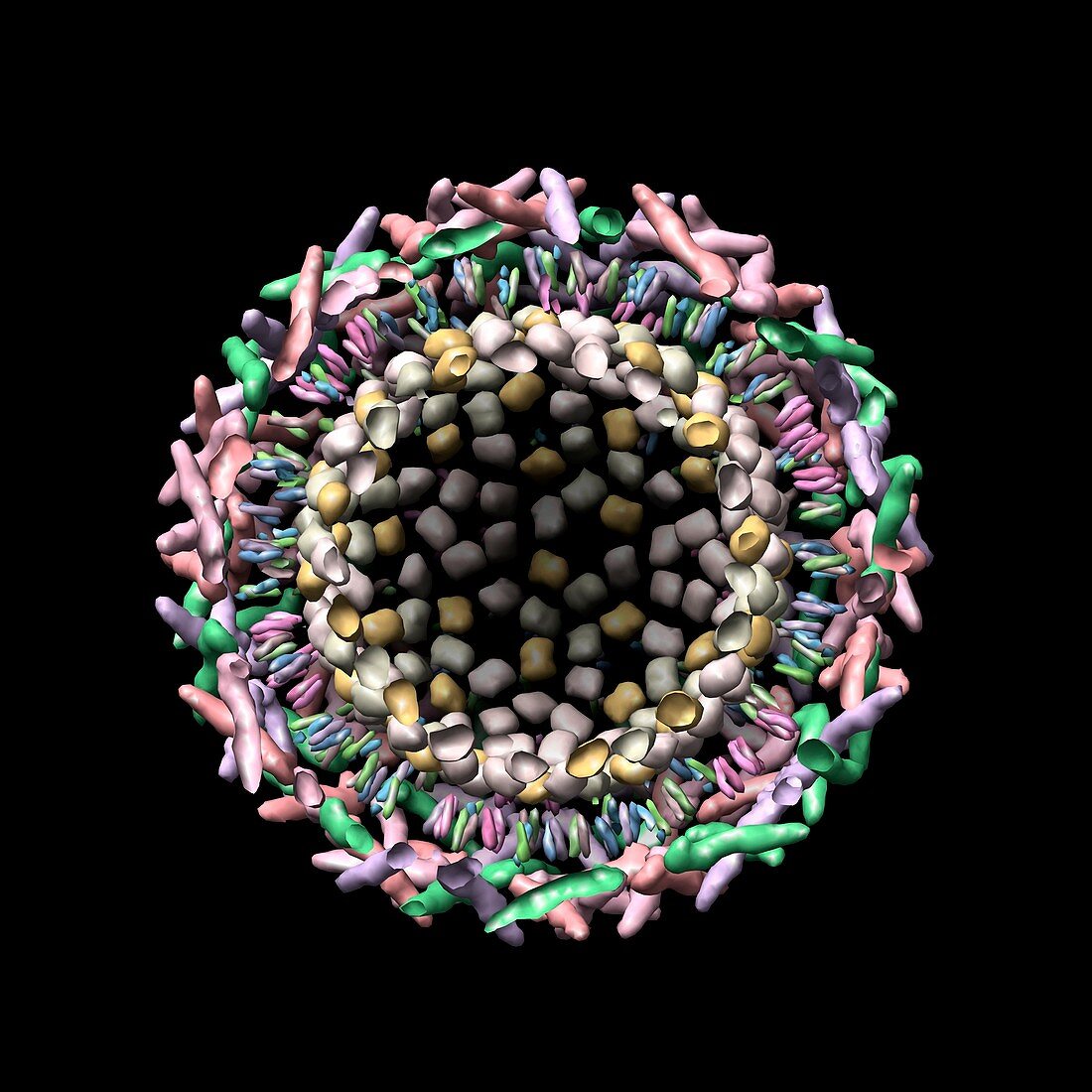
| Sindbis virus,computer model. Cross section of sindbis virus created using UCSF Chimera molecular modelling software and data from cryo-electron microscopy. It shows the outer glycoprotein shell and the inner protein shell (known as the capsid) separated by a lipid bilayer. The interior of the shell normally contains ribonucleic acid (RNA). Sindbis virus is transmitted by mosquitoes and can cause fever and rash. Cryo-electron microscopes fire beams of electrons at multiple angles at specimens kept at minus 150 degrees Celsius. The resulting 'slices' of data are reconstructed by programs,such as UCSF Chimera,into 3-D models | |
| Lizenzart: | Lizenzpflichtig |
| Credit: | Science Photo Library / UCSF Chimera |
| Bildgröße: | 2965 px × 2965 px |
| Modell-Rechte: | nicht erforderlich |
| Eigentums-Rechte: | nicht erforderlich |
| Restrictions: | - |
Preise für dieses Bild ab 15 €
Universitäten & Organisationen
(Informationsmaterial Digital, Informationsmaterial Print, Lehrmaterial Digital etc.)
ab 15 €
Redaktionell
(Bücher, Bücher: Sach- und Fachliteratur, Digitale Medien (redaktionell) etc.)
ab 30 €
Werbung
(Anzeigen, Aussenwerbung, Digitale Medien, Fernsehwerbung, Karten, Werbemittel, Zeitschriften etc.)
ab 55 €
Handelsprodukte
(bedruckte Textilie, Kalender, Postkarte, Grußkarte, Verpackung etc.)
ab 75 €
Pauschalpreise
Rechtepakete für die unbeschränkte Bildnutzung in Print oder Online
ab 495 €
Keywords
- 3-d,
- 3D,
- Biologie,
- biologisch,
- cgi,
- Chimäre,
- Cryo-EM,
- Cutaway,
- digital generiert,
- Dreidimensional,
- Eiweiß,
- Elektronenmikroskopie,
- Erreger,
- Gestalt,
- Illustration,
- Kapsid,
- Kryo-EM,
- Kryoelektronenmikroskopie,
- Kunstwerk,
- Makromolekül,
- Medizin,
- medizinisch,
- Mikrobiologie,
- mikrobiologisch,
- Modelling,
- molekulare Struktur,
- Molekülmodell,
- Oberfläche,
- Partikel,
- PDB,
- Querschnitt,
- Sindbis-Virus,
- Struktur,
- viral,
- Viren,
- Virion,
- Virologie,
- Virus,
- Wiederaufbau