Scanning electron microscope
Bildnummer 11832547
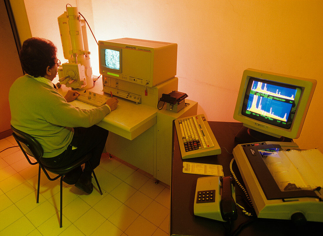
| Scanning electron microscope. Researcher looking at a sample using a scanning electron microscope (SEM). An SEM uses an electron beam to obtain a three-dimensional image of an object. The beam is scanned over the sample in a vacuum,causing the emission of secondary electrons. These secondary electrons are detected and used to form the image. The sample here is material from a fossilised footprint found in rock on the Roccamoufina Volcano,near Naples,Italy. It is part of a set known locally as the 'Devil's tracks'. The footprints have been identified as human and dated to between 385,000 and 325,00 years ago. Photographed at the University of Naples,Italy | |
| Lizenzart: | Lizenzpflichtig |
| Credit: | Science Photo Library / Sorrentino, Pasquale |
| Bildgröße: | 4893 px × 3582 px |
| Modell-Rechte: | Derzeit liegt noch kein Release vor. Bitte kontaktieren Sie uns vor Verwendung. |
| Eigentums-Rechte: | nicht erforderlich |
| Restrictions: |
|
Preise für dieses Bild ab 15 €
Universitäten & Organisationen
(Informationsmaterial Digital, Informationsmaterial Print, Lehrmaterial Digital etc.)
ab 15 €
Redaktionell
(Bücher, Bücher: Sach- und Fachliteratur, Digitale Medien (redaktionell) etc.)
ab 30 €
Werbung
(Anzeigen, Aussenwerbung, Digitale Medien, Fernsehwerbung, Karten, Werbemittel, Zeitschriften etc.)
ab 55 €
Handelsprodukte
(bedruckte Textilie, Kalender, Postkarte, Grußkarte, Verpackung etc.)
ab 75 €
Pauschalpreise
Rechtepakete für die unbeschränkte Bildnutzung in Print oder Online
ab 495 €
Keywords
- 50er Jahre,
- Analyse,
- Ausrüstung,
- Chemie,
- chemisch,
- Erwachsene,
- Europa,
- europäisch,
- Forscher,
- Fossil,
- Fünfziger Jahre,
- Gerät,
- Instrument,
- Italien,
- Italienisch,
- kaukasisch,
- Labor,
- Mann,
- Männlich,
- Maschine,
- Mensch,
- Monitor,
- Muster,
- Probe,
- Rasterelektronenmikroskop,
- rasterelektronenmikroskopische Aufnahme,
- REM,
- Technologie,
- technologisch,
- Teufelsspuren,
- vorbereitend,
- weiß,
- Wissenschaft,
- Wissenschaftler,
- wissenschaftlich