Scanning electron microscopy
Bildnummer 11832544
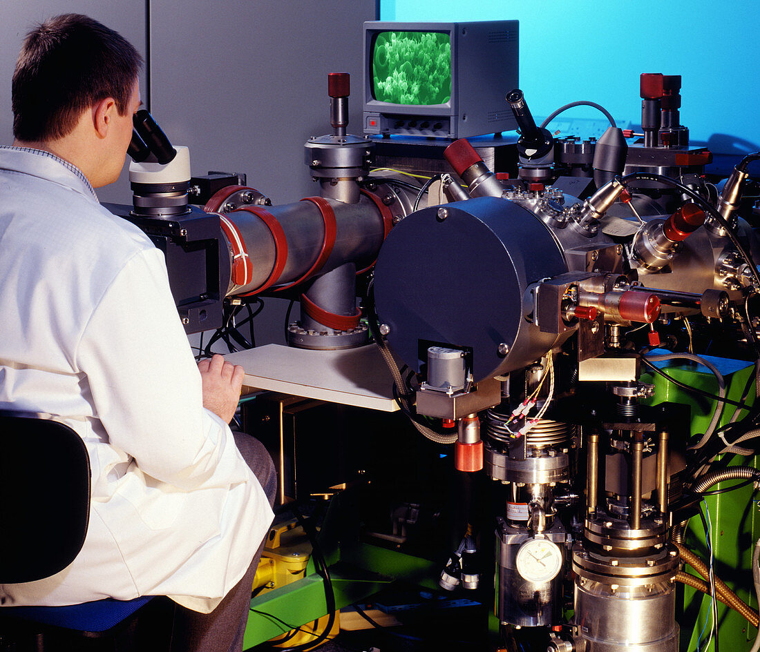
| Scanning electron microscope. Researcher using a scanning electron microscope (SEM,upper left) to view blood cells. SEMs use an electron beam to obtain three-dimensional images of objects. The electron beam is moved across the sample,which causes secondary electrons to be emitted. It is these electrons which are used to form the image. SEMs can provide magnifications as high as 300,000 times original size. Photographed in the UK | |
| Lizenzart: | Lizenzpflichtig |
| Credit: | Science Photo Library / Allen, Steve |
| Bildgröße: | 4492 px × 3860 px |
| Modell-Rechte: | Derzeit liegt noch kein Release vor. Bitte kontaktieren Sie uns vor Verwendung. |
| Eigentums-Rechte: | nicht erforderlich |
| Restrictions: | - |
Preise für dieses Bild ab 15 €
Universitäten & Organisationen
(Informationsmaterial Digital, Informationsmaterial Print, Lehrmaterial Digital etc.)
ab 15 €
Redaktionell
(Bücher, Bücher: Sach- und Fachliteratur, Digitale Medien (redaktionell) etc.)
ab 30 €
Werbung
(Anzeigen, Aussenwerbung, Digitale Medien, Fernsehwerbung, Karten, Werbemittel, Zeitschriften etc.)
ab 55 €
Handelsprodukte
(bedruckte Textilie, Kalender, Postkarte, Grußkarte, Verpackung etc.)
ab 75 €
Pauschalpreise
Rechtepakete für die unbeschränkte Bildnutzung in Print oder Online
ab 495 €
Keywords
- Anzeige,
- anzeigen,
- Arbeit,
- Arbeiten,
- Ausrüstung,
- Bildschirm,
- Blut,
- britisch,
- forschend,
- Forscher,
- Gerät,
- Großbritannien,
- Instrument,
- Mann,
- Männlich,
- Maschine,
- Mikroskopie,
- Monitor,
- Nutzung,
- Physik,
- Rasterelektronenmikroskop,
- REM,
- rot,
- schauend,
- Technik,
- Techniken,
- Techniker,
- Technologie,
- technologisch,
- Vereinigtes Königreich,
- Wissenschaft,
- Wissenschaftler,
- wissenschaftlich,
- Zelle,
- Zellen