HeLa cancer cells
Bildnummer 11821007
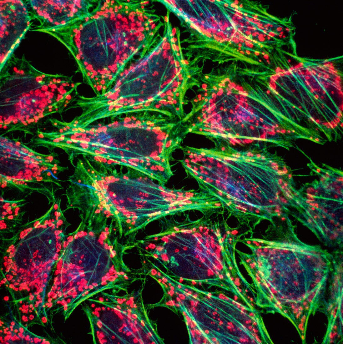
| HeLa cancer cells. Confocal light micrograph of HeLa cancer cells in culture. The cells have been stained with fluorescent markers to show the cell nucleus (blue),energy-producing mitochondria (pink) and fibres of the structural protein actin (green). HeLa cells are widely used in biological research because they can be grown indefinitely in culture. They were the first human cell line to be established and were derived from a cervical carcinoma sample taken from Henrietta Lacks in 1926. Confocal light microscopy uses light to excite fluorescent dye molecules bound to specific proteins in the cell. Magnification unknown | |
| Lizenzart: | Lizenzpflichtig |
| Credit: | Science Photo Library / Murti, Dr. Gopal |
| Bildgröße: | 3739 px × 3749 px |
| Modell-Rechte: | nicht erforderlich |
| Eigentums-Rechte: | nicht erforderlich |
| Restrictions: | - |
Preise für dieses Bild ab 15 €
Universitäten & Organisationen
(Informationsmaterial Digital, Informationsmaterial Print, Lehrmaterial Digital etc.)
ab 15 €
Redaktionell
(Bücher, Bücher: Sach- und Fachliteratur, Digitale Medien (redaktionell) etc.)
ab 30 €
Werbung
(Anzeigen, Aussenwerbung, Digitale Medien, Fernsehwerbung, Karten, Werbemittel, Zeitschriften etc.)
ab 55 €
Handelsprodukte
(bedruckte Textilie, Kalender, Postkarte, Grußkarte, Verpackung etc.)
ab 75 €
Pauschalpreise
Rechtepakete für die unbeschränkte Bildnutzung in Print oder Online
ab 495 €