F/col STM image of DNA (isometric projection)
Bildnummer 11817722
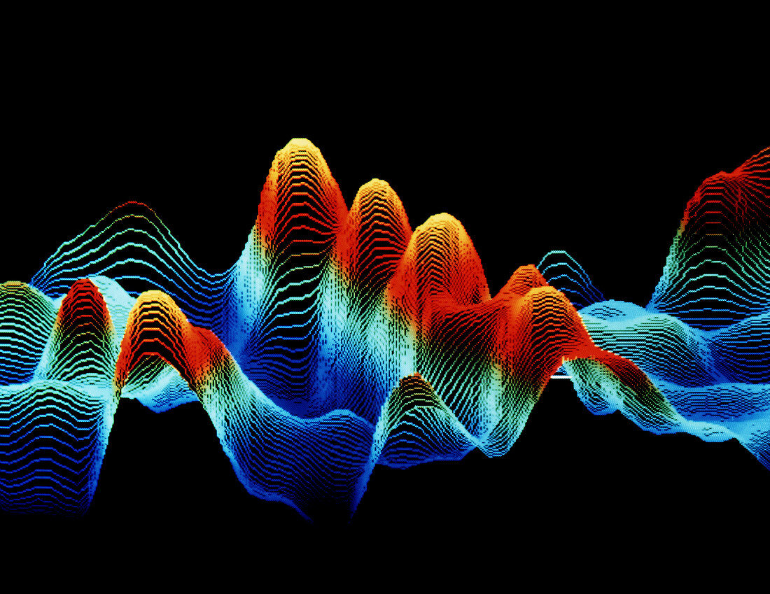
| False-colour scanning tunnelling micrograph (STM) of DNA. A sample of uncoated,double-stranded DNA was dissolved in a salt solution & deposited on graphite prior to being imaged in air by the STM. An STM image is formed by scanning a fine point just above the specimen surface & electronically recording the height of the point as it moves. This image shows a section of a double-stranded DNA molecule (a DNA duplex),with the coils of the helix appearing as the row of orange/yellow peaks in the centre of the image. The average distance between each peak is 3.5 nanometers. Magnification: x1,500,000 at 6x7cm size | |
| Lizenzart: | Lizenzpflichtig |
| Credit: | Science Photo Library / Lawrence Berkeley Laboratory |
| Bildgröße: | 4126 px × 3184 px |
| Modell-Rechte: | nicht erforderlich |
| Eigentums-Rechte: | nicht erforderlich |
| Restrictions: | - |
Preise für dieses Bild ab 15 €
Universitäten & Organisationen
(Informationsmaterial Digital, Informationsmaterial Print, Lehrmaterial Digital etc.)
ab 15 €
Redaktionell
(Bücher, Bücher: Sach- und Fachliteratur, Digitale Medien (redaktionell) etc.)
ab 30 €
Werbung
(Anzeigen, Aussenwerbung, Digitale Medien, Fernsehwerbung, Karten, Werbemittel, Zeitschriften etc.)
ab 55 €
Handelsprodukte
(bedruckte Textilie, Kalender, Postkarte, Grußkarte, Verpackung etc.)
ab 75 €
Pauschalpreise
Rechtepakete für die unbeschränkte Bildnutzung in Print oder Online
ab 495 €