Molar tooth cross-section,artwork
Bildnummer 11814085
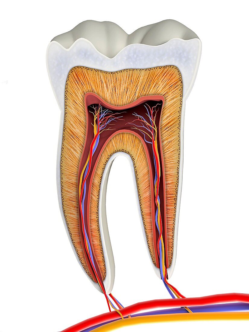
| Molar tooth cross-section,artwork. The upper (biting) surfaces of the tooth are at top,with the lower sections (bottom) embedded in the gums and jaw bone (not shown). The cross-section shows the tooth's internal anatomy,including the living tissue of the pulp (red) in the tooth's core and its roots (bottom). Within the pulp are venous (blue) and arterial (red) capillaries (small blood vessels),as well as nerves (orange). It is inflammation and infection of this living tissue that causes the pain of a toothache if tooth decay causes loss of the overlying inorganic enamel and dentine layers | |
| Lizenzart: | Lizenzfrei |
| Credit: | Science Photo Library / Pasieka, Alfred |
| Modell-Rechte: | nicht erforderlich |
| Eigentums-Rechte: | nicht erforderlich |
| Restrictions: | - |
Preise für dieses Bild ab 29 €
Für digitale Nutzung (72 dpi)
ab 29 €
Für Druckauflösung (300 dpi)
ab 300 €
Keywords
- 3D,
- Anatomie,
- anatomisch,
- anorganisch,
- Arterie,
- arteriell,
- arterielle Kapillaren,
- Arterien,
- ausgeschnitten,
- Ausschnitte,
- Biologie,
- biologisch,
- Blutgefäß,
- Blutgefäße,
- Cutaway,
- dental,
- Dentin,
- einer,
- einfacher Hintergrund,
- Emaille,
- gesund,
- Gesundheit,
- Gesundheitswesen,
- Gewebe,
- Illustration,
- kapillar,
- Kreislauf,
- Kunstwerk,
- lebendes Gewebe,
- menschlicher Körper,
- Mineral,
- Mund,
- Nerv,
- Nerven,
- Nervensystem,
- neural,
- Niemand,
- normal,
- Organisch,
- Physiologie,
- physiologisch,
- Querschnitt,
- Schicht,
- Schichten,
- Sektion,
- sektioniert,
- Single,
- vaskulär,
- Vene,
- Venen,
- venös,
- venöse Kapillaren,
- weißer Hintergrund,
- Wurzeln,
- Zahn,
- Zähne