Arsenite-stressed cells,light micrograph
Bildnummer 11734332
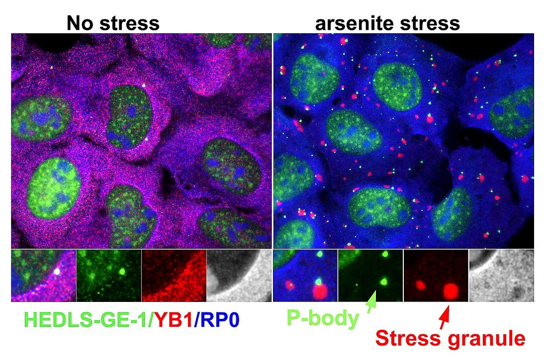
| Arsenite-stressed cells. Immunofluorescence light micrograph of cells stained to show cell proteins and stress responses to arsenite. The image at left shows no stress,with the image at right showing stress due to arsenite (chemical compounds containing arsenic). The text at lower left gives the names of the proteins and the colours of their corresponding stains: HEDLS-GE-1 (green),YB1 (red,Y box binding protein 1),and RP0 (blue,ribosomal P0 protein). At right are inset images showing P-bodies (green,processing bodies containing RNA enzymes) and stress granules (red) | |
| Lizenzart: | Lizenzpflichtig |
| Credit: | Science Photo Library / Kedersha, Nancy |
| Bildgröße: | 5126 px × 3416 px |
| Modell-Rechte: | nicht erforderlich |
| Eigentums-Rechte: | nicht erforderlich |
| Restrictions: | - |
Preise für dieses Bild ab 15 €
Universitäten & Organisationen
(Informationsmaterial Digital, Informationsmaterial Print, Lehrmaterial Digital etc.)
ab 15 €
Redaktionell
(Bücher, Bücher: Sach- und Fachliteratur, Digitale Medien (redaktionell) etc.)
ab 30 €
Werbung
(Anzeigen, Aussenwerbung, Digitale Medien, Fernsehwerbung, Karten, Werbemittel, Zeitschriften etc.)
ab 55 €
Handelsprodukte
(bedruckte Textilie, Kalender, Postkarte, Grußkarte, Verpackung etc.)
ab 75 €
Pauschalpreise
Rechtepakete für die unbeschränkte Bildnutzung in Print oder Online
ab 495 €