Human ear anatomy,illustration
Bildnummer 11729974
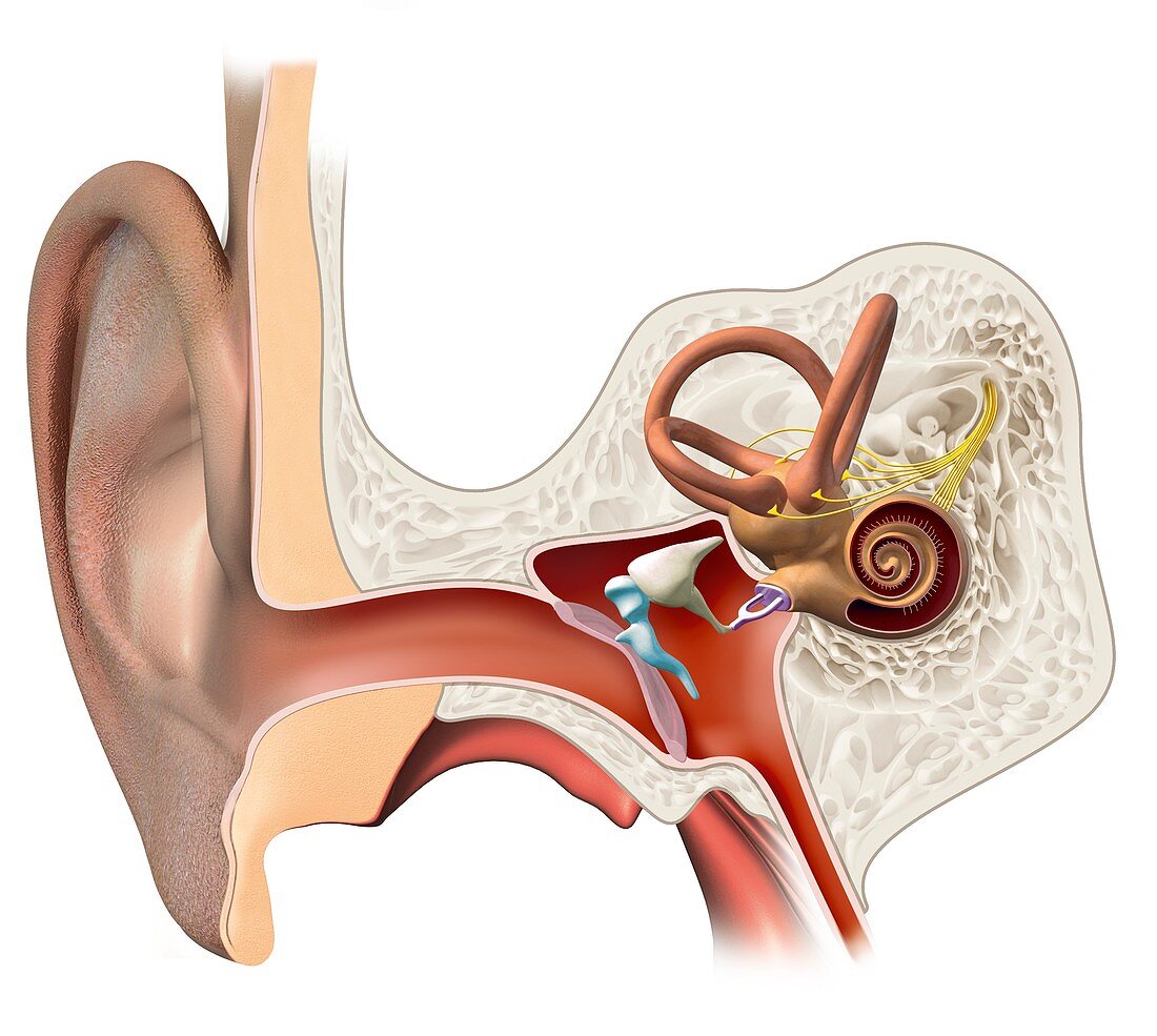
| Human ear anatomy. Computer illustration of a section through a human ear,the organ of hearing and balance. The ear canal (centre left) of the outer ear (left) joins the pinna (the visible part of the ear) to the eardrum (pink,centre),the start of the middle ear. Three tiny joined bones (the ossicles,centre-right): the malleus,joined to the inside of the eardrum,the incus,and the stapes,connect the eardrum to the inner ear (right). The inner ear consists of fluid-filled passages called the labyrinth,which includes the snail-like cochlea (curled,right) that is connected by nerves (yellow) to the brain. The loops above the cochlea are semi-circular canals responsible for balance. The Eustachian tube (lower right) joins the middle ear to the nasopharynx | |
| Lizenzart: | Lizenzpflichtig |
| Credit: | Science Photo Library / Lunau, Claus |
| Bildgröße: | 5315 px × 4759 px |
| Modell-Rechte: | nicht erforderlich |
| Eigentums-Rechte: | nicht erforderlich |
| Restrictions: | - |
Preise für dieses Bild ab 15 €
Universitäten & Organisationen
(Informationsmaterial Digital, Informationsmaterial Print, Lehrmaterial Digital etc.)
ab 15 €
Redaktionell
(Bücher, Bücher: Sach- und Fachliteratur, Digitale Medien (redaktionell) etc.)
ab 30 €
Werbung
(Anzeigen, Aussenwerbung, Digitale Medien, Fernsehwerbung, Karten, Werbemittel, Zeitschriften etc.)
ab 55 €
Handelsprodukte
(bedruckte Textilie, Kalender, Postkarte, Grußkarte, Verpackung etc.)
ab 75 €
Pauschalpreise
Rechtepakete für die unbeschränkte Bildnutzung in Print oder Online
ab 495 €