Fetal testis,light micrograph
Bildnummer 11723353
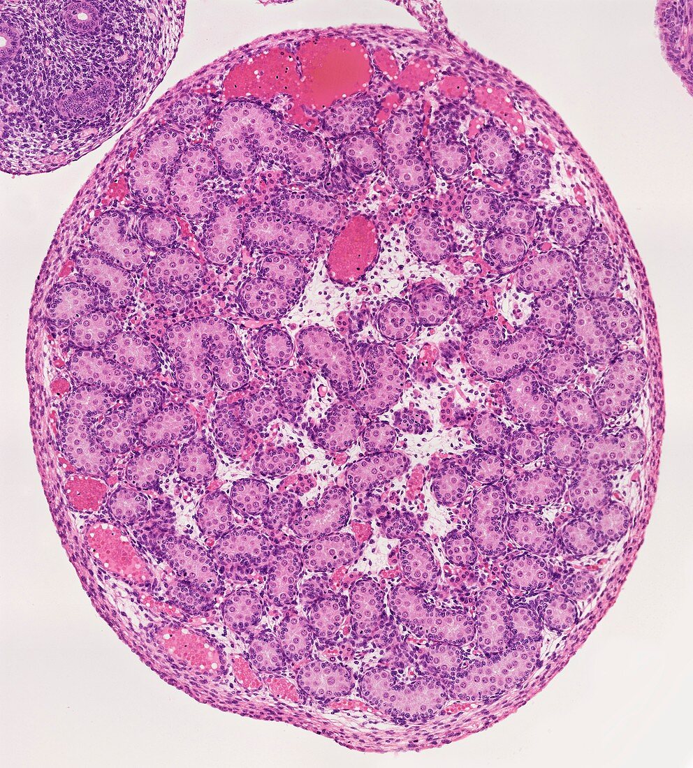
| Light microscopy of a fetal testis. The characteristic features are the solid,coiled seminiferous cords that in postnatal life will become seminiferous tubules,and clusters of Leydig cells (red) between the cords. The cords contain Sertoli cells and germ cells termed spermatogonia (round cells). The spermatogonia will become the stem cells for subsequent sperm development during spermatogenesis. Blood vessels some large,are located just deep to the testis capsule. Magnification x80 when printed at 10 cm wide | |
| Lizenzart: | Lizenzpflichtig |
| Credit: | Science Photo Library / Microscape |
| Bildgröße: | 4048 px × 4488 px |
| Modell-Rechte: | nicht erforderlich |
| Eigentums-Rechte: | nicht erforderlich |
| Restrictions: | - |
Preise für dieses Bild ab 15 €
Universitäten & Organisationen
(Informationsmaterial Digital, Informationsmaterial Print, Lehrmaterial Digital etc.)
ab 15 €
Redaktionell
(Bücher, Bücher: Sach- und Fachliteratur, Digitale Medien (redaktionell) etc.)
ab 30 €
Werbung
(Anzeigen, Aussenwerbung, Digitale Medien, Fernsehwerbung, Karten, Werbemittel, Zeitschriften etc.)
ab 55 €
Handelsprodukte
(bedruckte Textilie, Kalender, Postkarte, Grußkarte, Verpackung etc.)
ab 75 €
Pauschalpreise
Rechtepakete für die unbeschränkte Bildnutzung in Print oder Online
ab 495 €