Nerve tissue,light micrograph
Bildnummer 11723317
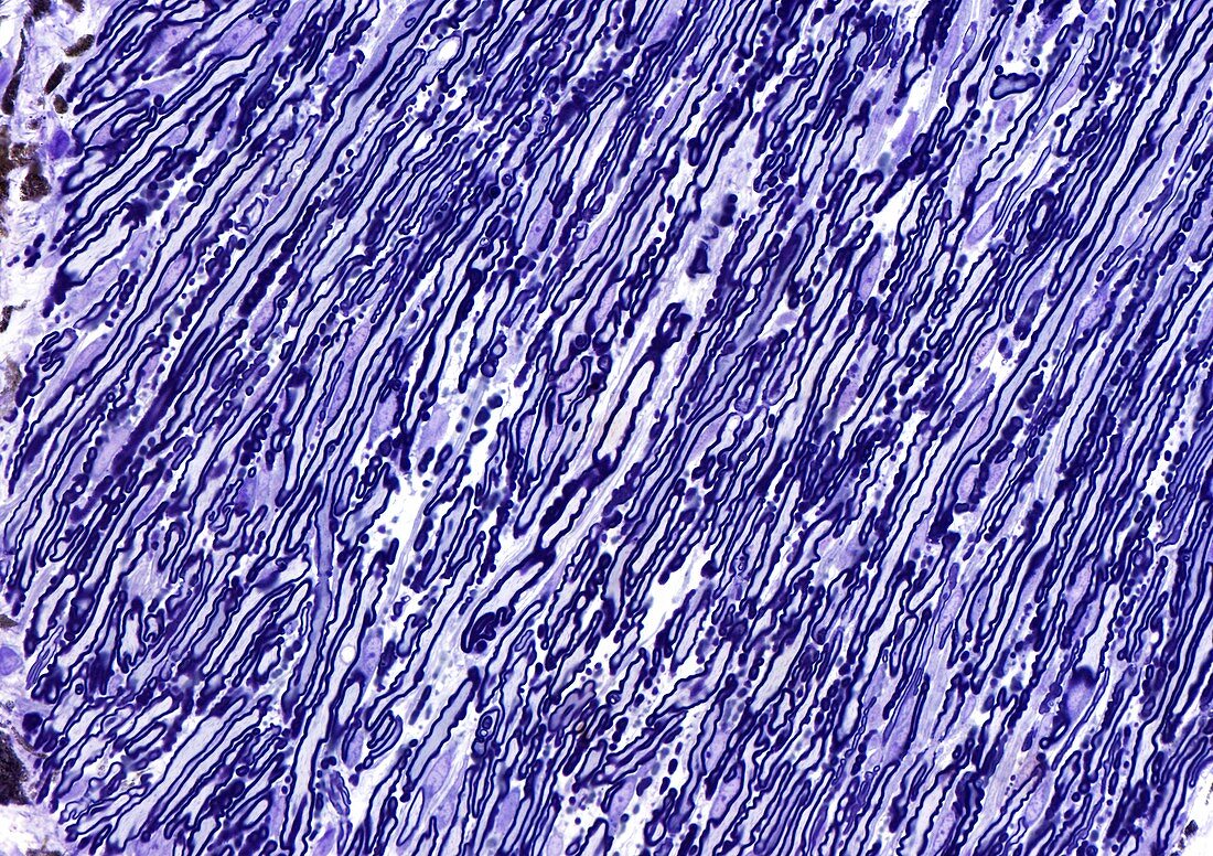
| Light microscopy of myelinated nerves in a toluidine blue-stained section of the optic nerve. Axons appear as mostly empty-looking and pale grey-blue elongated profiles enclosed by deeply stained margins representing myelin sheaths produced by Schwann cells. Slender flattened nuclei between nerve fibres are the Schwann cell nuclei. There are about one million axons in each optic nerve. Magnification x550 when printed at 10 cm height | |
| Lizenzart: | Lizenzpflichtig |
| Credit: | Science Photo Library / Microscape |
| Bildgröße: | 4961 px × 3499 px |
| Modell-Rechte: | nicht erforderlich |
| Eigentums-Rechte: | nicht erforderlich |
| Restrictions: | - |
Preise für dieses Bild ab 15 €
Universitäten & Organisationen
(Informationsmaterial Digital, Informationsmaterial Print, Lehrmaterial Digital etc.)
ab 15 €
Redaktionell
(Bücher, Bücher: Sach- und Fachliteratur, Digitale Medien (redaktionell) etc.)
ab 30 €
Werbung
(Anzeigen, Aussenwerbung, Digitale Medien, Fernsehwerbung, Karten, Werbemittel, Zeitschriften etc.)
ab 55 €
Handelsprodukte
(bedruckte Textilie, Kalender, Postkarte, Grußkarte, Verpackung etc.)
ab 75 €
Pauschalpreise
Rechtepakete für die unbeschränkte Bildnutzung in Print oder Online
ab 495 €