Horse's skull,3D CT scan
Bildnummer 11715021
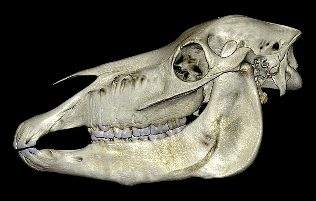
| Horse's skull. 3D computed tomography (CT) scan of the skull of a horse (Equus caballus). The skull is seen from the side. At far left right are the incisor teeth. Behind these is the interdental space,with the premolar and molar teeth at centre left and centre. An eye socket is at upper right,with the temporomandibular joint (TMJ) to its right. For this skull showing the pulp of the teeth,see image C025/4076. For this skull imaged from below by CT scanning,see image C025/4077 | |
| Lizenzart: | Lizenzpflichtig |
| Credit: | Science Photo Library / Persson, Anders / CMIV |
| Bildgröße: | 3741 px × 2374 px |
| Modell-Rechte: | nicht erforderlich |
| Eigentums-Rechte: | nicht erforderlich |
| Restrictions: | - |
Preise für dieses Bild ab 15 €
Universitäten & Organisationen
(Informationsmaterial Digital, Informationsmaterial Print, Lehrmaterial Digital etc.)
ab 15 €
Redaktionell
(Bücher, Bücher: Sach- und Fachliteratur, Digitale Medien (redaktionell) etc.)
ab 30 €
Werbung
(Anzeigen, Aussenwerbung, Digitale Medien, Fernsehwerbung, Karten, Werbemittel, Zeitschriften etc.)
ab 55 €
Handelsprodukte
(bedruckte Textilie, Kalender, Postkarte, Grußkarte, Verpackung etc.)
ab 75 €
Pauschalpreise
Rechtepakete für die unbeschränkte Bildnutzung in Print oder Online
ab 495 €
Keywords
- 3-d,
- 3D,
- Anatomie,
- anatomisch,
- Augenhöhle,
- ausgeschnitten,
- Ausschnitte,
- Biologie,
- biologisch,
- Computertomographie,
- CT-Scanner,
- Dreidimensional,
- Fauna,
- Gelenk,
- gesund,
- Joint,
- Kiefer,
- Knochen,
- Natur,
- Niemand,
- normal,
- Oberkiefer,
- Pferd,
- Pferde-,
- Prämolar,
- Probe,
- Profil,
- Säugetier,
- Schädel,
- Schläfenbein,
- Schneidezahn,
- schwarzer Hintergrund,
- Seitenansicht,
- seitlich,
- Tier,
- Tierwelt,
- Unterkiefer,
- Zahn,
- Zähne,
- Zoologie,
- zoologisch