Himalayan cedar (Cedrus deodara) leaf
Bildnummer 11712640
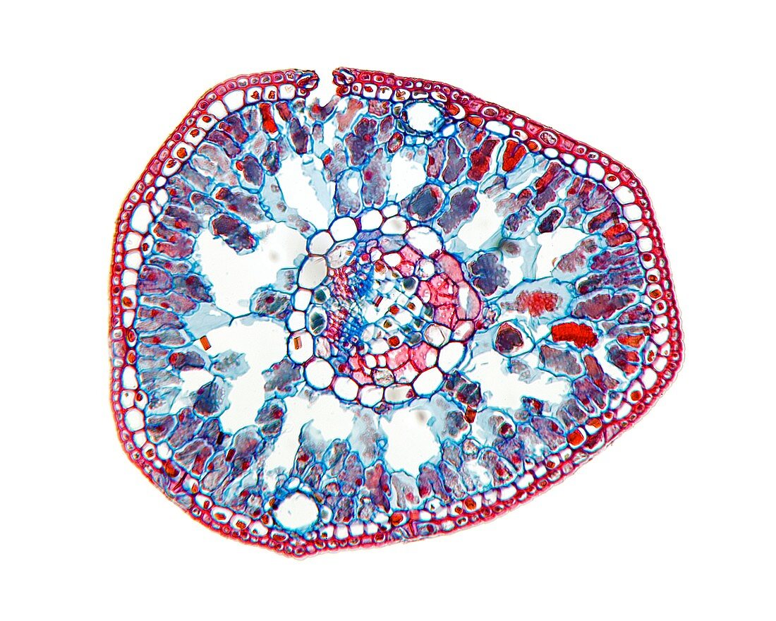
| Himalayan cedar (Cedrus deodara) leaf. Light micrograph of a section through a needle (leaf) from a Himalayan cedar tree,showing its thick cuticle and thick-walled epidermis (red) and hypodermis (subcutaneous tissue,red). Stomata (pores) and guard cells can also be seen,along with chloroplasts,and mesophyll tissue composed of parenchyma cells (blue-red). The vascular tissue is composed of xylem tracheid cells (red) and phloem sieve tube cells (blue). Magnification: x37 when printed 10 centimetres wide | |
| Lizenzart: | Lizenzpflichtig |
| Credit: | Science Photo Library / Wheeler, Dr. Keith |
| Bildgröße: | 4680 px × 3744 px |
| Modell-Rechte: | nicht erforderlich |
| Eigentums-Rechte: | nicht erforderlich |
| Restrictions: | - |
Preise für dieses Bild ab 15 €
Universitäten & Organisationen
(Informationsmaterial Digital, Informationsmaterial Print, Lehrmaterial Digital etc.)
ab 15 €
Redaktionell
(Bücher, Bücher: Sach- und Fachliteratur, Digitale Medien (redaktionell) etc.)
ab 30 €
Werbung
(Anzeigen, Aussenwerbung, Digitale Medien, Fernsehwerbung, Karten, Werbemittel, Zeitschriften etc.)
ab 55 €
Handelsprodukte
(bedruckte Textilie, Kalender, Postkarte, Grußkarte, Verpackung etc.)
ab 75 €
Pauschalpreise
Rechtepakete für die unbeschränkte Bildnutzung in Print oder Online
ab 495 €
Keywords
- Anatomie,
- anatomisch,
- ausgeschnitten,
- Ausschnitte,
- Baum,
- Biologie,
- biologisch,
- Blatt,
- Botanik,
- botanisch,
- Chloroplasten,
- epidermal,
- Epidermis,
- Flora,
- Gefäßband,
- Gewebe,
- gymnospermen,
- Himalaya-Zeder,
- Histologie,
- histologisch,
- Lichtmikroskop,
- lichtmikroskopische Aufnahme,
- Nacktsamer,
- Nadel,
- Nadelbaum,
- Natur,
- Niemand,
- Parenchym,
- Pflanze,
- Pflanzen,
- Phloem,
- Pore,
- Sektion,
- sektioniert,
- Stoma,
- Stomata,
- Tierwelt,
- weißer Hintergrund,
- Xylem,
- zapfentragend,
- Zelle,
- Zellen