Scots pine (Pinus sylvestris) leaf
Bildnummer 11712638
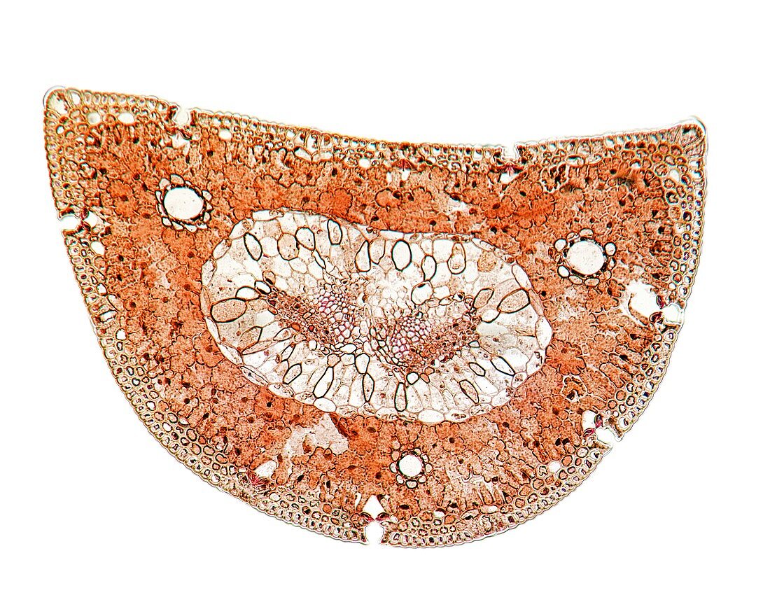
| Scots pine (Pinus sylvestris) leaf. Light micrograph of a section through a needle (leaf) from a Scots pine tree,showing its thick cuticle and thick-walled epidermis (brown) and hypodermis (subcutaneous tissue,brown). Stomata (pores) and guard cells can also be seen,along with chloroplasts,and mesophyll tissue composed of parenchyma cells. The vascular tissue is composed of xylem tracheid cells (pink) and phloem sieve tube cells (brown). Magnification: x37 when printed 10 centimetres wide | |
| Lizenzart: | Lizenzpflichtig |
| Credit: | Science Photo Library / Wheeler, Dr. Keith |
| Bildgröße: | 4902 px × 3921 px |
| Modell-Rechte: | nicht erforderlich |
| Eigentums-Rechte: | nicht erforderlich |
| Restrictions: | - |
Preise für dieses Bild ab 15 €
Universitäten & Organisationen
(Informationsmaterial Digital, Informationsmaterial Print, Lehrmaterial Digital etc.)
ab 15 €
Redaktionell
(Bücher, Bücher: Sach- und Fachliteratur, Digitale Medien (redaktionell) etc.)
ab 30 €
Werbung
(Anzeigen, Aussenwerbung, Digitale Medien, Fernsehwerbung, Karten, Werbemittel, Zeitschriften etc.)
ab 55 €
Handelsprodukte
(bedruckte Textilie, Kalender, Postkarte, Grußkarte, Verpackung etc.)
ab 75 €
Pauschalpreise
Rechtepakete für die unbeschränkte Bildnutzung in Print oder Online
ab 495 €
Keywords
- Anatomie,
- anatomisch,
- ausgeschnitten,
- Ausschnitte,
- Baum,
- Biologie,
- biologisch,
- Blatt,
- Botanik,
- botanisch,
- Chloroplasten,
- epidermal,
- Epidermis,
- Flora,
- Gefäßband,
- Gewebe,
- gymnospermen,
- Histologie,
- histologisch,
- Lichtmikroskop,
- lichtmikroskopische Aufnahme,
- Nacktsamer,
- Nadel,
- Nadelbaum,
- Natur,
- Niemand,
- Parenchym,
- Pflanze,
- Pflanzen,
- Phloem,
- Pore,
- Sektion,
- sektioniert,
- Stoma,
- Stomata,
- Tierwelt,
- weißer Hintergrund,
- Xylem,
- zapfentragend,
- Zelle,
- Zellen