Zebrafish eye,confocal micrograph
Bildnummer 11710257
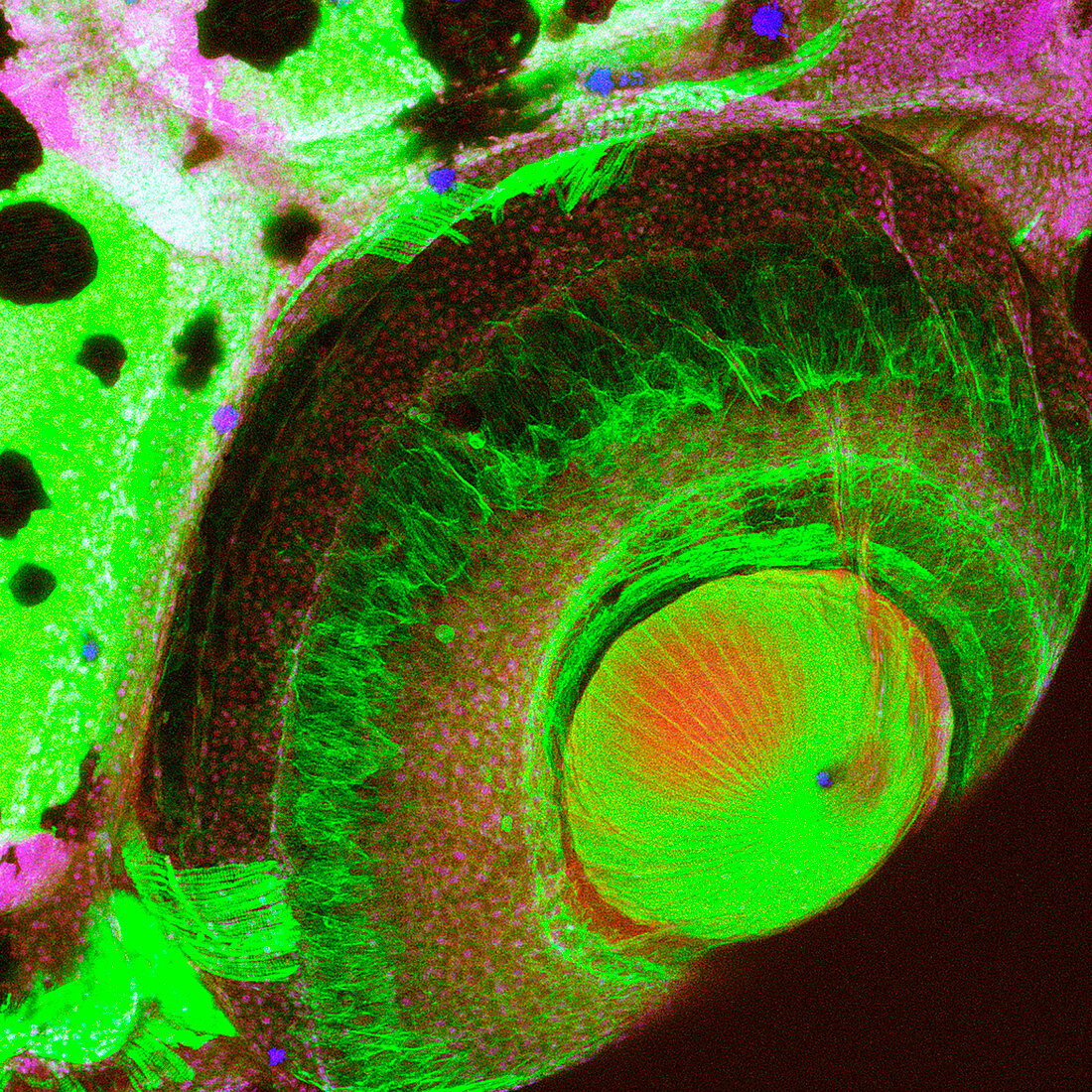
| Zebrafish eye. Scanning confocal light micrograph of tissue from the eye of a zebrafish,showing the lens (round,lower right). The image has been stained for cytoskeletal fibres (green). The staining and autofluorescence has also highlighted other proteins and DNA. This image includes the zonular fibres (bottom left) that connect the lens to the ciliary muscle. This image consists of about 20 optical sections as a collapsed stack. The zebrafish (Danio rerio) is a popular model organism in biological research. Magnification: x165 when printed at 10 centimetres across | |
| Lizenzart: | Lizenzpflichtig |
| Credit: | Science Photo Library |
| Bildgröße: | 4179 px × 4179 px |
| Modell-Rechte: | nicht erforderlich |
| Eigentums-Rechte: | nicht erforderlich |
| Restrictions: | - |
Preise für dieses Bild ab 15 €
Universitäten & Organisationen
(Informationsmaterial Digital, Informationsmaterial Print, Lehrmaterial Digital etc.)
ab 15 €
Redaktionell
(Bücher, Bücher: Sach- und Fachliteratur, Digitale Medien (redaktionell) etc.)
ab 30 €
Werbung
(Anzeigen, Aussenwerbung, Digitale Medien, Fernsehwerbung, Karten, Werbemittel, Zeitschriften etc.)
ab 55 €
Handelsprodukte
(bedruckte Textilie, Kalender, Postkarte, Grußkarte, Verpackung etc.)
ab 75 €
Pauschalpreise
Rechtepakete für die unbeschränkte Bildnutzung in Print oder Online
ab 495 €
Keywords
- Anatomie,
- anatomisch,
- Auge,
- Augenheilkunde,
- Autofluoreszenz,
- befleckt,
- Biologie,
- biologisch,
- Fauna,
- Fisch,
- Fluoreszenz,
- fluoreszierend,
- Gewebe,
- Histologie,
- histologisch,
- konfokal,
- Laser,
- Lichtmikroskop,
- lichtmikroskopische Aufnahme,
- Linse,
- Natur,
- Niemand,
- okular,
- optisch,
- Physiologie,
- physiologisch,
- Tier,
- Tierkörper,
- Tierwelt,
- Zoologie,
- zoologisch,
- Zytoskelett