Bone joint space,light micrograph
Bildnummer 11704724
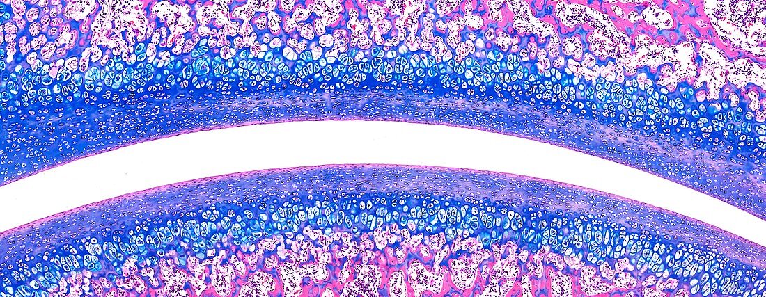
| Light microscopy of tissues covering the surfaces of two bone ends inside a joint. The opposing bone surfaces are covered with hyaline cartilage consisting of a matrix and cartilage cells (chondrocytes) stained blue. Beneath the cartilage zone is spongy bone (pink and blue) with gaps for bone marrow. The joint space is filled with synovial fluid acting as a lubricant and nutrient source. The cartilage has no direct blood vessels. Magnification x80 when narrow width printed at 10 cm | |
| Lizenzart: | Lizenzpflichtig |
| Credit: | Science Photo Library / Microscape |
| Bildgröße: | 6732 px × 2616 px |
| Modell-Rechte: | nicht erforderlich |
| Eigentums-Rechte: | nicht erforderlich |
| Restrictions: | - |
Preise für dieses Bild ab 15 €
Universitäten & Organisationen
(Informationsmaterial Digital, Informationsmaterial Print, Lehrmaterial Digital etc.)
ab 15 €
Redaktionell
(Bücher, Bücher: Sach- und Fachliteratur, Digitale Medien (redaktionell) etc.)
ab 30 €
Werbung
(Anzeigen, Aussenwerbung, Digitale Medien, Fernsehwerbung, Karten, Werbemittel, Zeitschriften etc.)
ab 55 €
Handelsprodukte
(bedruckte Textilie, Kalender, Postkarte, Grußkarte, Verpackung etc.)
ab 75 €
Pauschalpreise
Rechtepakete für die unbeschränkte Bildnutzung in Print oder Online
ab 495 €