Lens of eye,light micrograph
Bildnummer 11704299
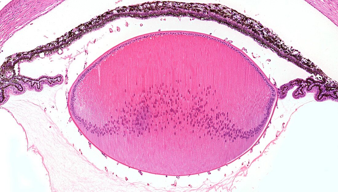
| Light microscopy of a section through the lens from the eye of a developing fetus. In the developing fetal lens the surface epithelial cells (top) multiply and become curved and elongated to form long lens fibres. The earliest formed lens fibres are located centrally,with new fibres added in layers at the periphery. As lens fibres mature they lose their nuclei and become transparent thereby contributing to focussing light onto the retina. Lens fibres are deformable,allowing the lens to change shape (a process known as accommodation) and allow focus on close or distant objects. Also noted is a section through the pigmented iris and ciliary bodies. Magnification x50 when printed at 10 cm | |
| Lizenzart: | Lizenzpflichtig |
| Credit: | Science Photo Library / Microscape |
| Bildgröße: | 5546 px × 3151 px |
| Modell-Rechte: | nicht erforderlich |
| Eigentums-Rechte: | nicht erforderlich |
| Restrictions: | - |
Preise für dieses Bild ab 15 €
Universitäten & Organisationen
(Informationsmaterial Digital, Informationsmaterial Print, Lehrmaterial Digital etc.)
ab 15 €
Redaktionell
(Bücher, Bücher: Sach- und Fachliteratur, Digitale Medien (redaktionell) etc.)
ab 30 €
Werbung
(Anzeigen, Aussenwerbung, Digitale Medien, Fernsehwerbung, Karten, Werbemittel, Zeitschriften etc.)
ab 55 €
Handelsprodukte
(bedruckte Textilie, Kalender, Postkarte, Grußkarte, Verpackung etc.)
ab 75 €
Pauschalpreise
Rechtepakete für die unbeschränkte Bildnutzung in Print oder Online
ab 495 €