Ovarian follicle,light micrograph
Bildnummer 11704285
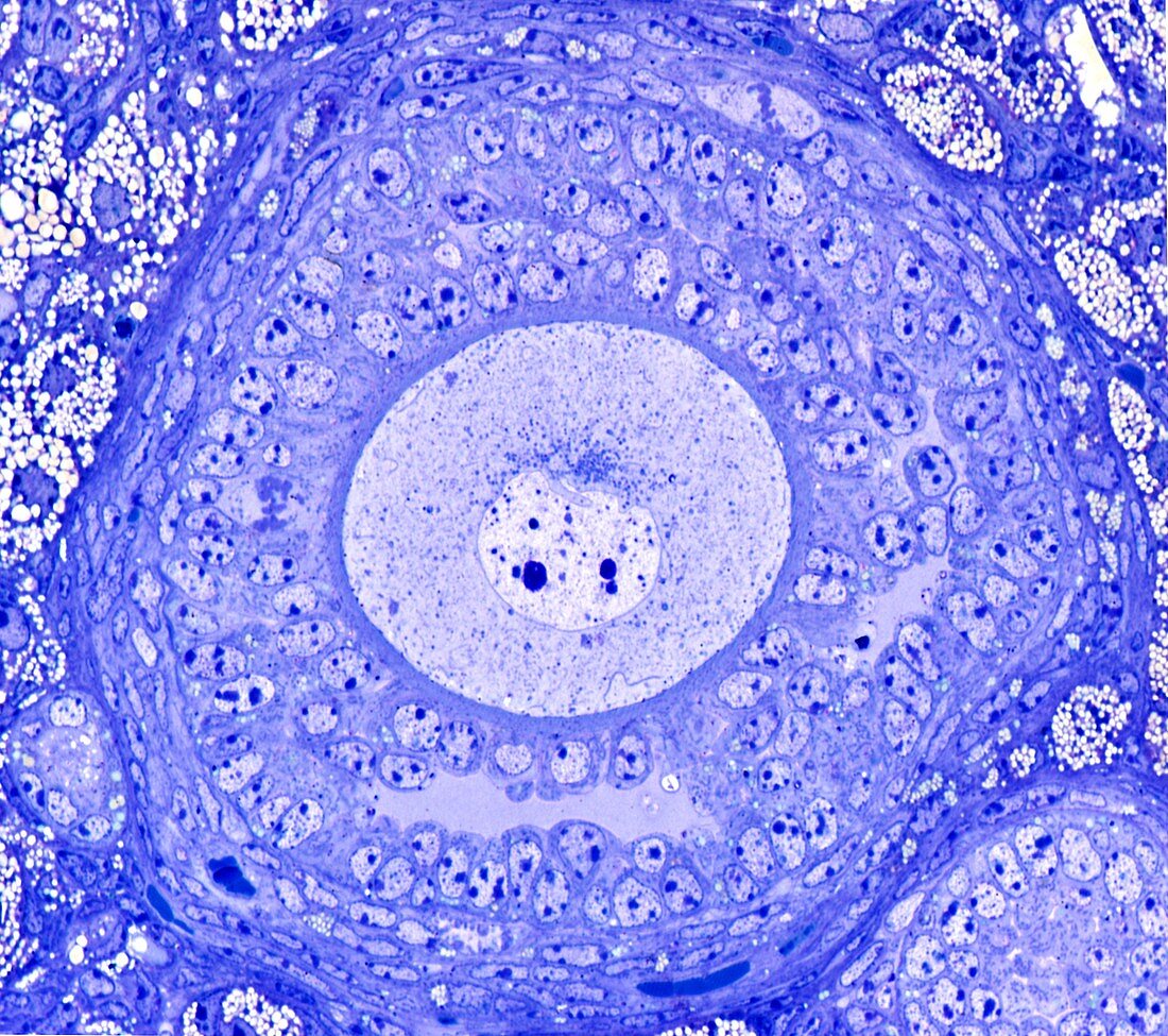
| Light microscopy of a growing ovarian follicle at the early antral stage of development. The central oocyte (female germ cell) is surrounded by follicular cells separated from the oocyte cytoplasm by a thin coat of special proteins called the zona pellucida which serves to bind sperm during fertilization,and to protect the early embryo if formed. Peripheral layers of flattened cells make up the thecal tissues. Fluid-filled gaps between follicular cells signify the initial stages of antrum formation during folliculogenesis which enlarge to dominate the volume of the follicle as it approaches ovulation. Magnification x500 when printed at 10 cm | |
| Lizenzart: | Lizenzpflichtig |
| Credit: | Science Photo Library / Microscape |
| Bildgröße: | 4439 px × 3936 px |
| Modell-Rechte: | nicht erforderlich |
| Eigentums-Rechte: | nicht erforderlich |
| Restrictions: | - |
Preise für dieses Bild ab 15 €
Universitäten & Organisationen
(Informationsmaterial Digital, Informationsmaterial Print, Lehrmaterial Digital etc.)
ab 15 €
Redaktionell
(Bücher, Bücher: Sach- und Fachliteratur, Digitale Medien (redaktionell) etc.)
ab 30 €
Werbung
(Anzeigen, Aussenwerbung, Digitale Medien, Fernsehwerbung, Karten, Werbemittel, Zeitschriften etc.)
ab 55 €
Handelsprodukte
(bedruckte Textilie, Kalender, Postkarte, Grußkarte, Verpackung etc.)
ab 75 €
Pauschalpreise
Rechtepakete für die unbeschränkte Bildnutzung in Print oder Online
ab 495 €