Growing bone,light micrograph
Bildnummer 11704284
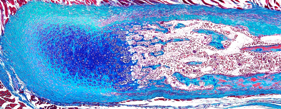
| Light microscopy of a growing rib bone. At one end the blue and green staining tissues are hyaline cartilage cells (chondrocytes) and matrix. Central cavities contain bone marrow. The opposite end shows red and green staining bone matrix making up the rib bone collar. The region where chondrocytes abut the bone marrow cavities is the epiphyseal growth plate. Here the chondrocytes die and their matrix is the site where newly forming bone is deposited by the process called endochondral ossification. Tissue margins around the cartilage are called perichondrium,that around the bone is called periosteum. Magnification x120 when narrow width printed at 10 cm | |
| Lizenzart: | Lizenzpflichtig |
| Credit: | Science Photo Library / Microscape |
| Bildgröße: | 6696 px × 2610 px |
| Modell-Rechte: | nicht erforderlich |
| Eigentums-Rechte: | nicht erforderlich |
| Restrictions: | - |
Preise für dieses Bild ab 15 €
Universitäten & Organisationen
(Informationsmaterial Digital, Informationsmaterial Print, Lehrmaterial Digital etc.)
ab 15 €
Redaktionell
(Bücher, Bücher: Sach- und Fachliteratur, Digitale Medien (redaktionell) etc.)
ab 30 €
Werbung
(Anzeigen, Aussenwerbung, Digitale Medien, Fernsehwerbung, Karten, Werbemittel, Zeitschriften etc.)
ab 55 €
Handelsprodukte
(bedruckte Textilie, Kalender, Postkarte, Grußkarte, Verpackung etc.)
ab 75 €
Pauschalpreise
Rechtepakete für die unbeschränkte Bildnutzung in Print oder Online
ab 495 €