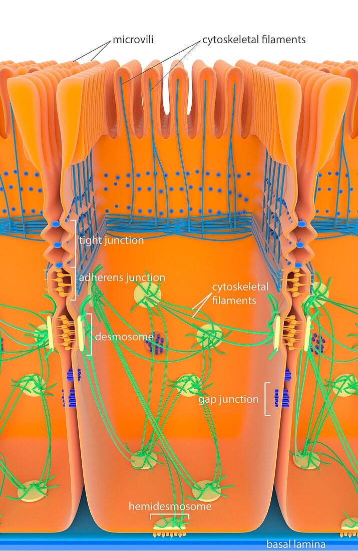
Intestinal cell junctions,illustration
Bildnummer 11703735

| Intestinal cell junctions. Illustration of an intestinal epithelial cell,showing anchoring junctions (desmosomes,yellow; adherens,orange),gap junctions (blue),tight junctions (ridged) and a hemidesmosome junction (bottom). Cytoskeletal filaments are blue and green. Across top are the microvilli that absorb nutrients from the intestinal lumen as food is digested. The various junctions shown here have a range of functions. Tight junctions form a relatively impermeable barrier. Anchoring junctions provide mechanical support. Gap junctions allows chemical and electrical communication between cells. For this artwork without labels,see C023/8828 | |
| Lizenzart: | Lizenzpflichtig |
| Credit: | Science Photo Library |
| Bildgröße: | 3403 px × 5238 px |
| Modell-Rechte: | nicht erforderlich |
| Eigentums-Rechte: | nicht erforderlich |
| Restrictions: | - |
Preise für dieses Bild ab 15 €
Universitäten & Organisationen
(Informationsmaterial Digital, Informationsmaterial Print, Lehrmaterial Digital etc.)
ab 15 €
Redaktionell
(Bücher, Bücher: Sach- und Fachliteratur, Digitale Medien (redaktionell) etc.)
ab 30 €
Werbung
(Anzeigen, Aussenwerbung, Digitale Medien, Fernsehwerbung, Karten, Werbemittel, Zeitschriften etc.)
ab 55 €
Handelsprodukte
(bedruckte Textilie, Kalender, Postkarte, Grußkarte, Verpackung etc.)
ab 75 €
Pauschalpreise
Rechtepakete für die unbeschränkte Bildnutzung in Print oder Online
ab 495 €
Keywords
- beschriftet,
- Biochemie,
- biochemisch,
- Biologie,
- biologisch,
- Darm,
- Darm-,
- Epithel,
- epithelial,
- Ernährung,
- Etikette,
- Etiketten,
- Gastroenterologie,
- Gedärme,
- Illustration,
- Kommunikation,
- Kunstwerk,
- Mikrovilli,
- Mikrovillus,
- Molekular,
- Niemand,
- Text,
- Verdauungskanal,
- Verdauungssystem,
- Verdauungstrakt,
- Zellbilogie,
- Zelle,
- zellular,
- Zytologie,
- Zytologisch,
- Zytoskelett
