Cell division during skin wound repair
Bildnummer 11703504
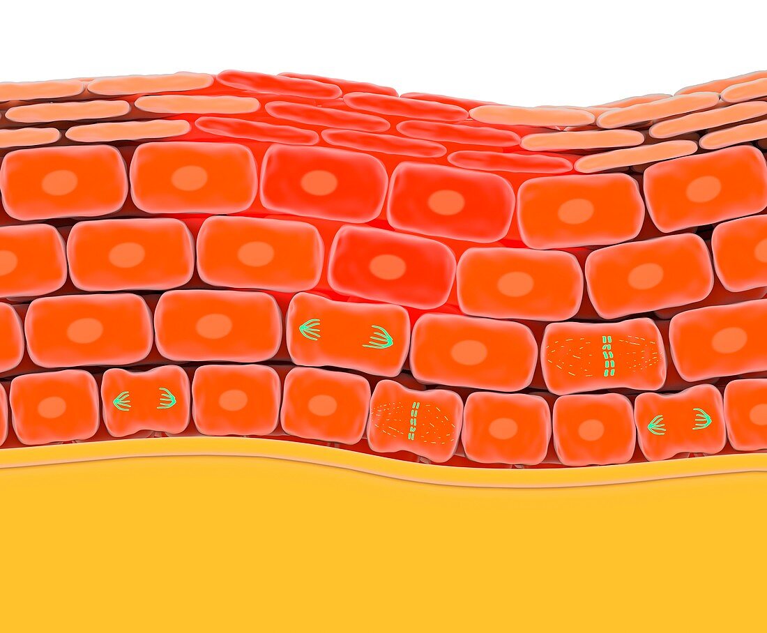
| Cell division during skin wound repair. Illustration of a vertical cross-section through skin,showing the cell layers and cell division in response to a wound (upper centre). Across bottom is connective tissue (yellow). The first layer of skin cells above the connective tissue is called the basal layer. Above that is the differentiation zone. At top is the outer layer of differentiated keratinised skin cells. Here,in addition to cell division (chromosomes,green) in the basal layer,cells are dividing around the wound site as part of the repair process. For a set of images showing cell division in normal,wounded and cancerous skin,see images C023/8583 to C023/8585 | |
| Lizenzart: | Lizenzpflichtig |
| Credit: | Science Photo Library |
| Bildgröße: | 4601 px × 3796 px |
| Modell-Rechte: | nicht erforderlich |
| Eigentums-Rechte: | nicht erforderlich |
| Restrictions: | - |
Preise für dieses Bild ab 15 €
Universitäten & Organisationen
(Informationsmaterial Digital, Informationsmaterial Print, Lehrmaterial Digital etc.)
ab 15 €
Redaktionell
(Bücher, Bücher: Sach- und Fachliteratur, Digitale Medien (redaktionell) etc.)
ab 30 €
Werbung
(Anzeigen, Aussenwerbung, Digitale Medien, Fernsehwerbung, Karten, Werbemittel, Zeitschriften etc.)
ab 55 €
Handelsprodukte
(bedruckte Textilie, Kalender, Postkarte, Grußkarte, Verpackung etc.)
ab 75 €
Pauschalpreise
Rechtepakete für die unbeschränkte Bildnutzung in Print oder Online
ab 495 €
Keywords
- Anatomie,
- anatomisch,
- Bindegewebe,
- Biologie,
- biologisch,
- Chromosomen,
- dermal,
- Dermatologie,
- dermatologisch,
- epidermal,
- Epidermis,
- Epithel,
- epithelial,
- gesund,
- Gewebe,
- Haut,
- Illustration,
- Keratin,
- Kunstwerk,
- menschlicher Körper,
- Niemand,
- normal,
- Physiologie,
- physiologisch,
- Querschnitt,
- Reihenfolge,
- Reparieren,
- Sektion,
- sektioniert,
- Serie,
- Teilen,
- weißer Hintergrund,
- Zelle,
- Zellen,
- zellular