Small intestine,light micrograph
Bildnummer 11703234
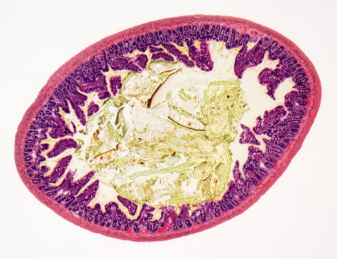
| Small intestine. Light micrograph of a section through the entire width of the small intestine. The small intestine is where digestion begins and nutrients are absorbed into the blood. The lumen (white,centre) is lined with villi (purple) and contains partially digested food (yellow). The folds in the intestinal surface greatly increase the surface area for absorption. The smooth muscle layer beneath the villi is used to mix the food as it passes through the lumen. The thick outer layer (pink) is composed of circular and longitudinal muscle tissue,the peristaltic action of which pushes the food along. Magnification: x60 when printed at 10cm wide | |
| Lizenzart: | Lizenzpflichtig |
| Credit: | Science Photo Library / Gschmeissner, Steve |
| Bildgröße: | 4769 px × 3664 px |
| Modell-Rechte: | nicht erforderlich |
| Eigentums-Rechte: | nicht erforderlich |
| Restrictions: | - |
Preise für dieses Bild ab 15 €
Universitäten & Organisationen
(Informationsmaterial Digital, Informationsmaterial Print, Lehrmaterial Digital etc.)
ab 15 €
Redaktionell
(Bücher, Bücher: Sach- und Fachliteratur, Digitale Medien (redaktionell) etc.)
ab 30 €
Werbung
(Anzeigen, Aussenwerbung, Digitale Medien, Fernsehwerbung, Karten, Werbemittel, Zeitschriften etc.)
ab 55 €
Handelsprodukte
(bedruckte Textilie, Kalender, Postkarte, Grußkarte, Verpackung etc.)
ab 75 €
Pauschalpreise
Rechtepakete für die unbeschränkte Bildnutzung in Print oder Online
ab 495 €
Keywords
- Absorption,
- Anatomie,
- anatomisch,
- befleckt,
- Biologie,
- biologisch,
- Darm,
- diagonal,
- Dünndarm,
- eingefärbt,
- farbig,
- Ganz,
- Ganze,
- gefärbt,
- gesund,
- Gewebe,
- Histologie,
- histologisch,
- Lichtmikroskop,
- lichtmikroskopische Aufnahme,
- Lumen,
- menschlicher Körper,
- Muscularis mucosae,
- Muskel,
- normal,
- Querschnitt,
- Scheibe,
- Schichten,
- Sektion,
- sektioniert,
- Verdauung,
- Verdauungssystem,
- Verfärbung,
- weiß,
- Zellen,
- Zotte,
- Zotten