Knee joint,light micrograph
Bildnummer 11703230
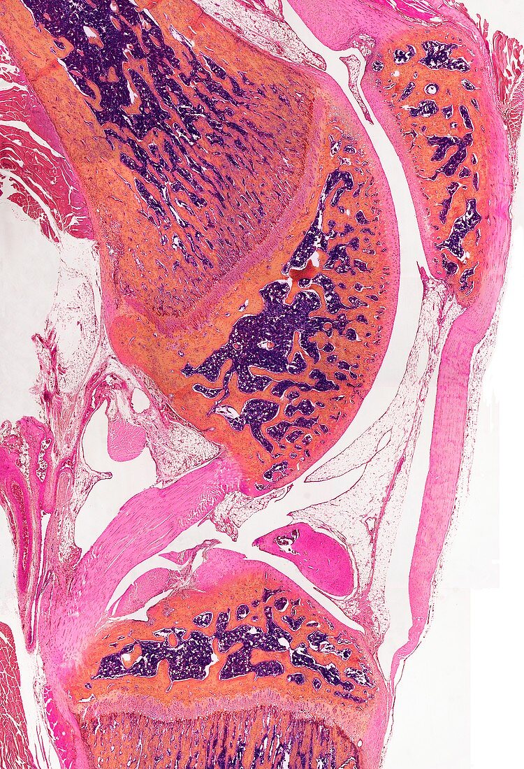
| Knee joint. Light micrograph of a section though a knee joint. The femur is to the top with the patella to the right. Anterior and posterior cruciate ligaments (pink) attach to the tibia below. The ends of the long bones are covered in articular cartilage that lines the synovial cavity. In osteoarthritis this cartilage becomes worn and rough causing inflammation and pain and may lead to the need for surgical knee replacement. Magnification: x5 when printed at 10 centimetres high | |
| Lizenzart: | Lizenzpflichtig |
| Credit: | Science Photo Library / Gschmeissner, Steve |
| Bildgröße: | 3451 px × 5064 px |
| Modell-Rechte: | nicht erforderlich |
| Eigentums-Rechte: | nicht erforderlich |
| Restrictions: | - |
Preise für dieses Bild ab 15 €
Universitäten & Organisationen
(Informationsmaterial Digital, Informationsmaterial Print, Lehrmaterial Digital etc.)
ab 15 €
Redaktionell
(Bücher, Bücher: Sach- und Fachliteratur, Digitale Medien (redaktionell) etc.)
ab 30 €
Werbung
(Anzeigen, Aussenwerbung, Digitale Medien, Fernsehwerbung, Karten, Werbemittel, Zeitschriften etc.)
ab 55 €
Handelsprodukte
(bedruckte Textilie, Kalender, Postkarte, Grußkarte, Verpackung etc.)
ab 75 €
Pauschalpreise
Rechtepakete für die unbeschränkte Bildnutzung in Print oder Online
ab 495 €