Myofibril muscle tissue,TEM
Bildnummer 11700137
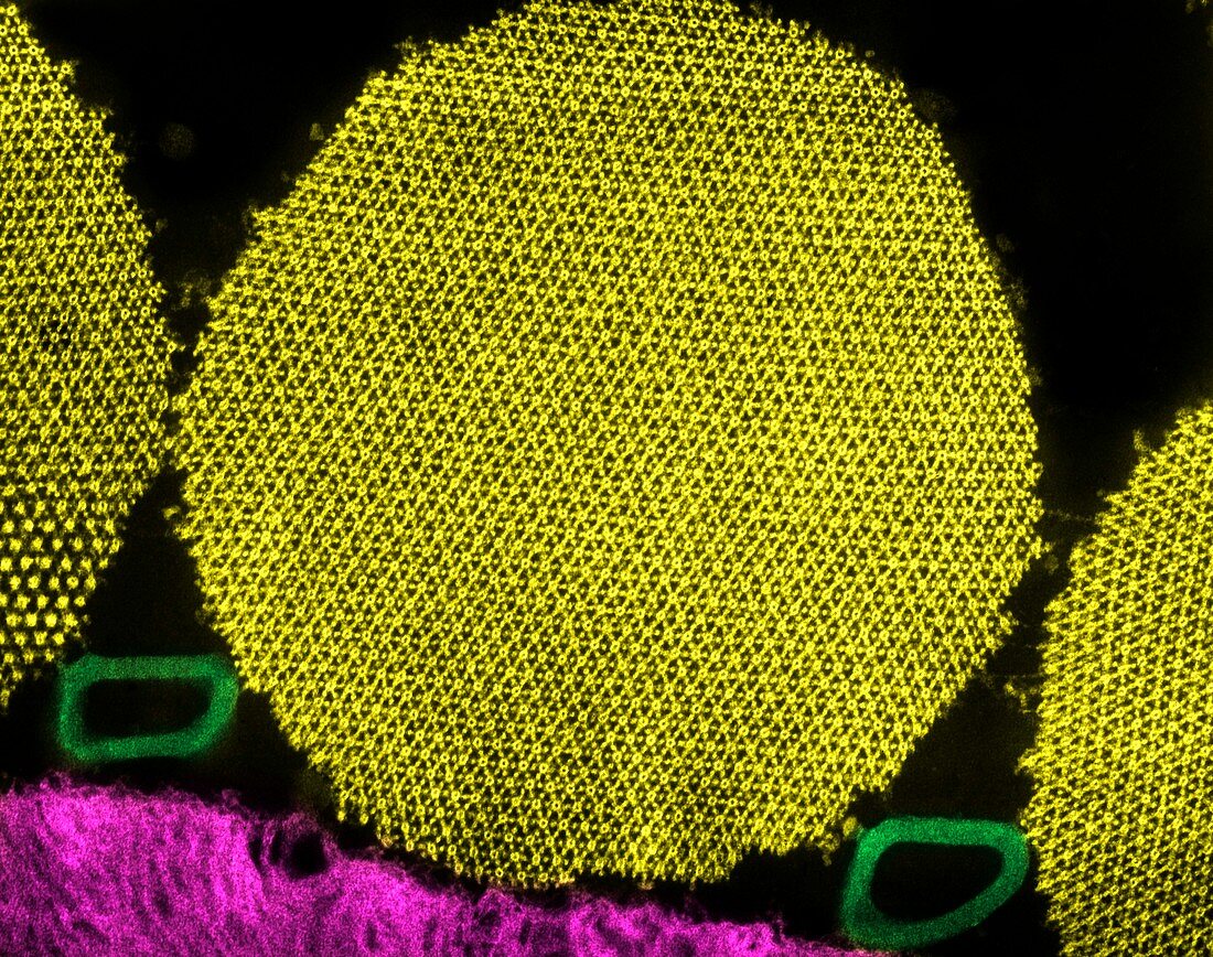
| Myofibril muscle tissue,coloured transmission electron micrograph (TEM). This cross-section through an insect flight muscle myofibril (round) shows the regular arrangement of the overlapping actin and myosin protein filaments. The larger circles are the myosin proteins and the smaller spots are the actin proteins. These proteins pull along each other to cause the muscle to contract. The myosin molecules shown here are around 50 nanometres apart. The rounded structures (green,lower left and lower right) between the myofibrils are tracheoles,which deliver oxygen to the muscles. Magnification: x35,500 when printed at 10 centimetres across | |
| Lizenzart: | Lizenzpflichtig |
| Credit: | Science Photo Library / AMMRF, UNIVERSITY OF SYDNEY |
| Bildgröße: | 4719 px × 3723 px |
| Modell-Rechte: | nicht erforderlich |
| Eigentums-Rechte: | nicht erforderlich |
| Restrictions: | - |
Preise für dieses Bild ab 15 €
Universitäten & Organisationen
(Informationsmaterial Digital, Informationsmaterial Print, Lehrmaterial Digital etc.)
ab 15 €
Redaktionell
(Bücher, Bücher: Sach- und Fachliteratur, Digitale Medien (redaktionell) etc.)
ab 30 €
Werbung
(Anzeigen, Aussenwerbung, Digitale Medien, Fernsehwerbung, Karten, Werbemittel, Zeitschriften etc.)
ab 55 €
Handelsprodukte
(bedruckte Textilie, Kalender, Postkarte, Grußkarte, Verpackung etc.)
ab 75 €
Pauschalpreise
Rechtepakete für die unbeschränkte Bildnutzung in Print oder Online
ab 495 €
Keywords
- Aktin,
- Anatomie,
- anatomisch,
- Biologie,
- biologisch,
- farbig,
- gefärbt,
- gesund,
- Gewebe,
- Insekt,
- Kontraktion,
- Mikroskop,
- Muskel,
- Muskeln,
- Myofilamente,
- Niemand,
- normal,
- Physiologie,
- physiologisch,
- Proteine,
- Querschnitt,
- Sektion,
- sektioniert,
- Skelettmuskulatur,
- tem,
- Tierkörper,
- Transmissionselektronen,
- transmissionselektronenmikroskopische Aufnahme,
- Ultrastruktur