Small intestine tissue,light micrograph
Bildnummer 11693618
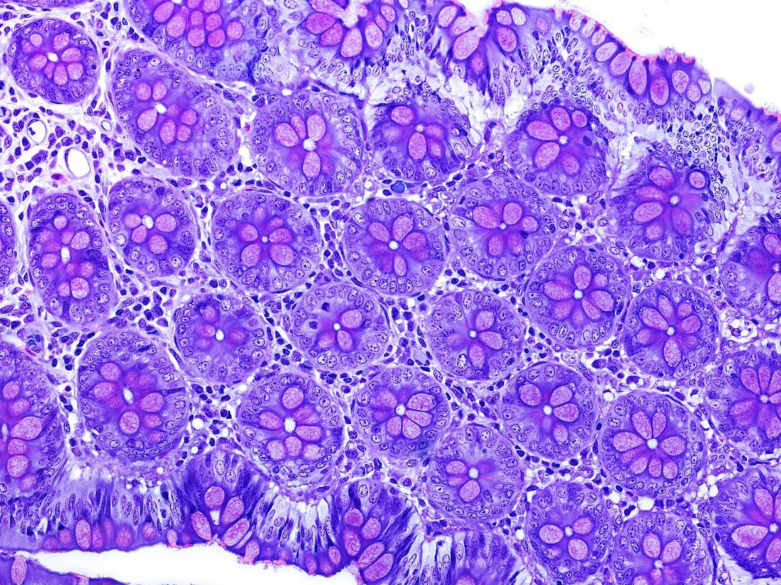
| Small intestine tissue. Light micrograph of a longitudinal section through tissue from the small intestine. This view shows cross-sections through many intestinal glands called crypts of Lieberkuhn. Crypts are long blind-ending tube-like extensions of the surface epithelial lining of the gut. In the small intestine they comprise several cell types including mucus-secreting goblet cells (purple) and absorptive enterocytes (blue) around a narrow central lumen. Crypts also contain gut epithelial stem cells. Connective tissue supporting the crypts contains fibroblasts,nerves,blood vessels,and white blood cells. Magnification: x183 when printed at 10 centimetres across | |
| Lizenzart: | Lizenzpflichtig |
| Credit: | Science Photo Library / Microscape |
| Bildgröße: | 4843 px × 3632 px |
| Modell-Rechte: | nicht erforderlich |
| Eigentums-Rechte: | nicht erforderlich |
| Restrictions: | - |
Preise für dieses Bild ab 15 €
Universitäten & Organisationen
(Informationsmaterial Digital, Informationsmaterial Print, Lehrmaterial Digital etc.)
ab 15 €
Redaktionell
(Bücher, Bücher: Sach- und Fachliteratur, Digitale Medien (redaktionell) etc.)
ab 30 €
Werbung
(Anzeigen, Aussenwerbung, Digitale Medien, Fernsehwerbung, Karten, Werbemittel, Zeitschriften etc.)
ab 55 €
Handelsprodukte
(bedruckte Textilie, Kalender, Postkarte, Grußkarte, Verpackung etc.)
ab 75 €
Pauschalpreise
Rechtepakete für die unbeschränkte Bildnutzung in Print oder Online
ab 495 €
Keywords
- Absorption,
- Anatomie,
- befleckt,
- Bindegewebe,
- Biologie,
- Darm,
- Drüse,
- Drüsen-,
- Dünndarm,
- Enterozyten,
- Epithel,
- epithelial,
- Gastroenterologie,
- gesund,
- Gewebe,
- Histologie,
- histologisch,
- Krypta,
- Krypten von Lieberkuhn,
- Lichtmikroskop,
- lichtmikroskopische Aufnahme,
- menschlicher Körper,
- Niemand,
- normal,
- Schleim,
- Sektion,
- sektioniert,
- Verdauung,
- Verdauungskanal,
- Verdauungssystem,
- Verdauungstrakt,
- Zelle,
- Zellen,
- zellular