Fields of vision,19th century artwork
Bildnummer 11692172
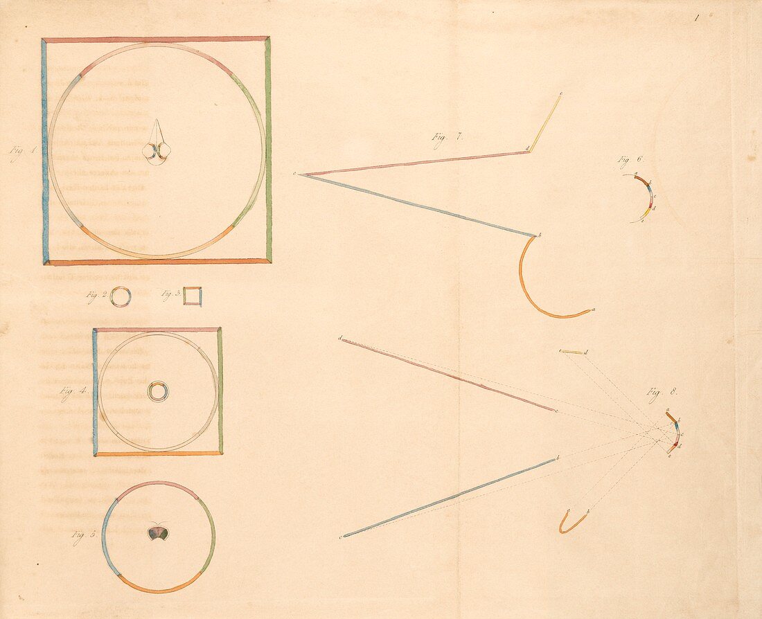
| Fields of vision. 19th century diagrams showing how the field of view maps to the retinas of different eyes. At top left is a bird's eye,at centre left is a simple eye and at bottom left is a fly's eye. At right the differences between a concave (top) and faceted (bottom) retina are shown. Artwork from 'Contributions to the Physiology of Vision' (1834) by the German physician Carl Moritz Nicolaus Bartels (1800-1835) | |
| Lizenzart: | Lizenzpflichtig |
| Credit: | Science Photo Library / King's College London |
| Bildgröße: | 4673 px × 3797 px |
| Modell-Rechte: | nicht erforderlich |
| Eigentums-Rechte: | nicht erforderlich |
| Restrictions: | - |
Preise für dieses Bild ab 15 €
Universitäten & Organisationen
(Informationsmaterial Digital, Informationsmaterial Print, Lehrmaterial Digital etc.)
ab 15 €
Redaktionell
(Bücher, Bücher: Sach- und Fachliteratur, Digitale Medien (redaktionell) etc.)
ab 30 €
Werbung
(Anzeigen, Aussenwerbung, Digitale Medien, Fernsehwerbung, Karten, Werbemittel, Zeitschriften etc.)
ab 55 €
Handelsprodukte
(bedruckte Textilie, Kalender, Postkarte, Grußkarte, Verpackung etc.)
ab 75 €
Pauschalpreise
Rechtepakete für die unbeschränkte Bildnutzung in Print oder Online
ab 495 €