Snail,micro-CT scan
Bildnummer 11686787
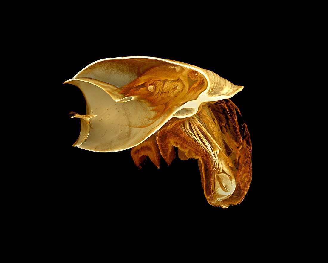
| Snail (Bostryx sp.),micro-CT scan,cross-section. This image was acquired using X-ray micro-Computed Tomography (also known as micro-CT). A micro-CT scanner projects a beam of X-rays through the sample onto a detector panel. Images are collected over a 360° rotation and these are then reconstructed to form a virtual 3D model of the specimen. This virtual object that can be viewed from all angles and sliced open or digitally dissected. This specimen was stained with iodine to provide contrast in the soft tissue,allowing features such as the gut,genitalia and muscles to be visualised. Image by Dan Sykes | |
| Lizenzart: | Lizenzpflichtig |
| Credit: | Science Photo Library / NATURAL HISTORY MUSEUM, LONDON / DAN SYKES |
| Bildgröße: | 3306 px × 2644 px |
| Modell-Rechte: | nicht erforderlich |
| Eigentums-Rechte: | nicht erforderlich |
| Restrictions: | - |
Preise für dieses Bild ab 15 €
Universitäten & Organisationen
(Informationsmaterial Digital, Informationsmaterial Print, Lehrmaterial Digital etc.)
ab 15 €
Redaktionell
(Bücher, Bücher: Sach- und Fachliteratur, Digitale Medien (redaktionell) etc.)
ab 30 €
Werbung
(Anzeigen, Aussenwerbung, Digitale Medien, Fernsehwerbung, Karten, Werbemittel, Zeitschriften etc.)
ab 55 €
Handelsprodukte
(bedruckte Textilie, Kalender, Postkarte, Grußkarte, Verpackung etc.)
ab 75 €
Pauschalpreise
Rechtepakete für die unbeschränkte Bildnutzung in Print oder Online
ab 495 €