Rhinoceros beetle,micro-CT scan
Bildnummer 11686741
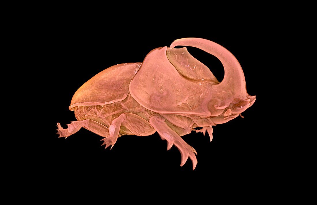
| Rhinoceros beetle (Oryctes boas),micro-CT scan. The micro-CT scanner was used to investigate the internal anatomy of this large beetle. This image was acquired using X-ray micro-Computed Tomography (also known as micro-CT). A micro-CT scanner projects a beam of X-rays through the sample onto a detector panel. Images are collected over a 360° rotation and these are then reconstructed to form a virtual 3D model of the specimen. This virtual object that can be viewed from all angles and sliced open or digitally dissected. Image by Dan Sykes | |
| Lizenzart: | Lizenzpflichtig |
| Credit: | Science Photo Library / NATURAL HISTORY MUSEUM, LONDON / DAN SYKES |
| Bildgröße: | 3694 px × 2386 px |
| Modell-Rechte: | nicht erforderlich |
| Eigentums-Rechte: | nicht erforderlich |
| Restrictions: | - |
Preise für dieses Bild ab 15 €
Universitäten & Organisationen
(Informationsmaterial Digital, Informationsmaterial Print, Lehrmaterial Digital etc.)
ab 15 €
Redaktionell
(Bücher, Bücher: Sach- und Fachliteratur, Digitale Medien (redaktionell) etc.)
ab 30 €
Werbung
(Anzeigen, Aussenwerbung, Digitale Medien, Fernsehwerbung, Karten, Werbemittel, Zeitschriften etc.)
ab 55 €
Handelsprodukte
(bedruckte Textilie, Kalender, Postkarte, Grußkarte, Verpackung etc.)
ab 75 €
Pauschalpreise
Rechtepakete für die unbeschränkte Bildnutzung in Print oder Online
ab 495 €