Double hip replacement,3D CT scan
Bildnummer 11682989
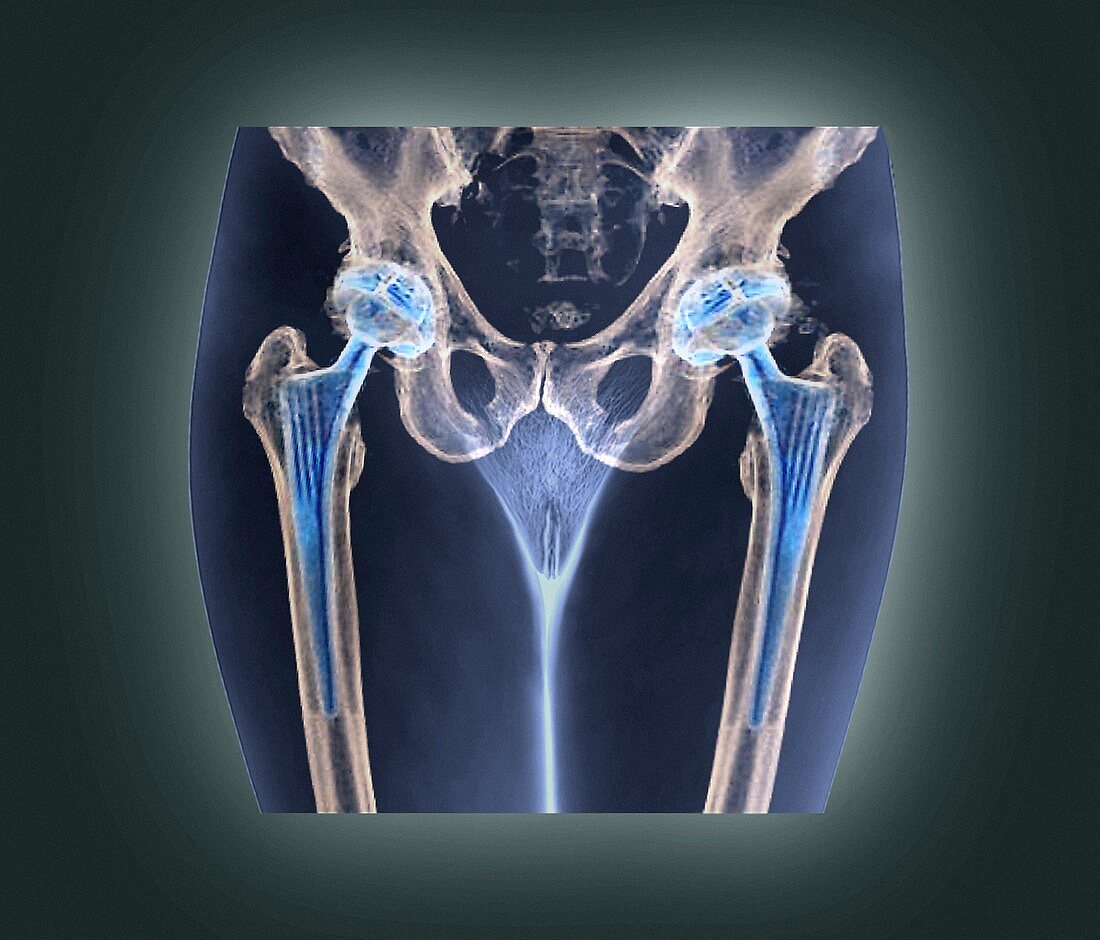
| Double hip replacement. Coloured 3D computed tomography (CT) scan of a 58 year old woman's pelvis (frontal view) with two total hip replacements (blue structures). These were implanted to treat osteoarthritis. Osteoarthritis is a condition where the cartilage cushioning the bones of the hip joint wears away. The bones then rub against each other,causing pain and stiffness. Surgery is required to replace the head of the femur (thigh bone) and acetabulum (hip joint socket) with a ball-and-shaft prosthesis | |
| Lizenzart: | Lizenzpflichtig |
| Credit: | Science Photo Library / Zephyr |
| Bildgröße: | 3206 px × 2740 px |
| Modell-Rechte: | nicht erforderlich |
| Eigentums-Rechte: | nicht erforderlich |
| Restrictions: | - |
Preise für dieses Bild ab 15 €
Universitäten & Organisationen
(Informationsmaterial Digital, Informationsmaterial Print, Lehrmaterial Digital etc.)
ab 15 €
Redaktionell
(Bücher, Bücher: Sach- und Fachliteratur, Digitale Medien (redaktionell) etc.)
ab 30 €
Werbung
(Anzeigen, Aussenwerbung, Digitale Medien, Fernsehwerbung, Karten, Werbemittel, Zeitschriften etc.)
ab 55 €
Handelsprodukte
(bedruckte Textilie, Kalender, Postkarte, Grußkarte, Verpackung etc.)
ab 75 €
Pauschalpreise
Rechtepakete für die unbeschränkte Bildnutzung in Print oder Online
ab 495 €