Brain tumour,fMRI
Bildnummer 11682960
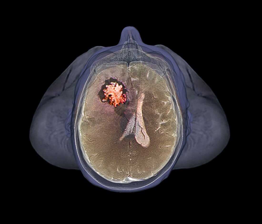
| Brain tumour,functional magnetic resonance imaging (fMRI) scan,from above. This is a 49 year old man with a brain lesion in the fronto-temporal cortex (top left),showing a grade 1 pilocytic astrocytoma (early stage,malignant brain tumour). This is a rare case,as pilocytic astrocytomas usually occur in young patients. The tumour has shifted the mid-line of the brain to the right. FMRI is used to detect changes in blood flow and blood oxygenation in the brain,highlighting areas of increased stimulation | |
| Lizenzart: | Lizenzpflichtig |
| Credit: | Science Photo Library / Zephyr |
| Bildgröße: | 4537 px × 3886 px |
| Modell-Rechte: | nicht erforderlich |
| Eigentums-Rechte: | nicht erforderlich |
| Restrictions: | - |
Preise für dieses Bild ab 15 €
Universitäten & Organisationen
(Informationsmaterial Digital, Informationsmaterial Print, Lehrmaterial Digital etc.)
ab 15 €
Redaktionell
(Bücher, Bücher: Sach- und Fachliteratur, Digitale Medien (redaktionell) etc.)
ab 30 €
Werbung
(Anzeigen, Aussenwerbung, Digitale Medien, Fernsehwerbung, Karten, Werbemittel, Zeitschriften etc.)
ab 55 €
Handelsprodukte
(bedruckte Textilie, Kalender, Postkarte, Grußkarte, Verpackung etc.)
ab 75 €
Pauschalpreise
Rechtepakete für die unbeschränkte Bildnutzung in Print oder Online
ab 495 €
Keywords
- 40er Jahre,
- abnormal,
- Aktivität,
- Bewegung,
- Bildgebung,
- Diagnose,
- Erwachsene,
- farbig,
- FMRI,
- funktionellen Magnetresonanztomographie,
- geduldig,
- gefärbt,
- Gehirn,
- Großhirn,
- Kondition,
- Krankheit,
- Krebs,
- linke Hemisphäre,
- maligne,
- Männlich,
- Medizin,
- medizinisch,
- Mensch,
- menschlicher Körper,
- MRT-Untersuchung,
- Neuroimaging,
- Neurologie,
- neurologisch,
- Schaden,
- schwarzer Hintergrund,
- Störung,
- Tumor,
- ungesund,
- Vierziger Jahre,
- Wunde,
- Zusammengesetzt