Brain tumour,fMRI scan
Bildnummer 11682959
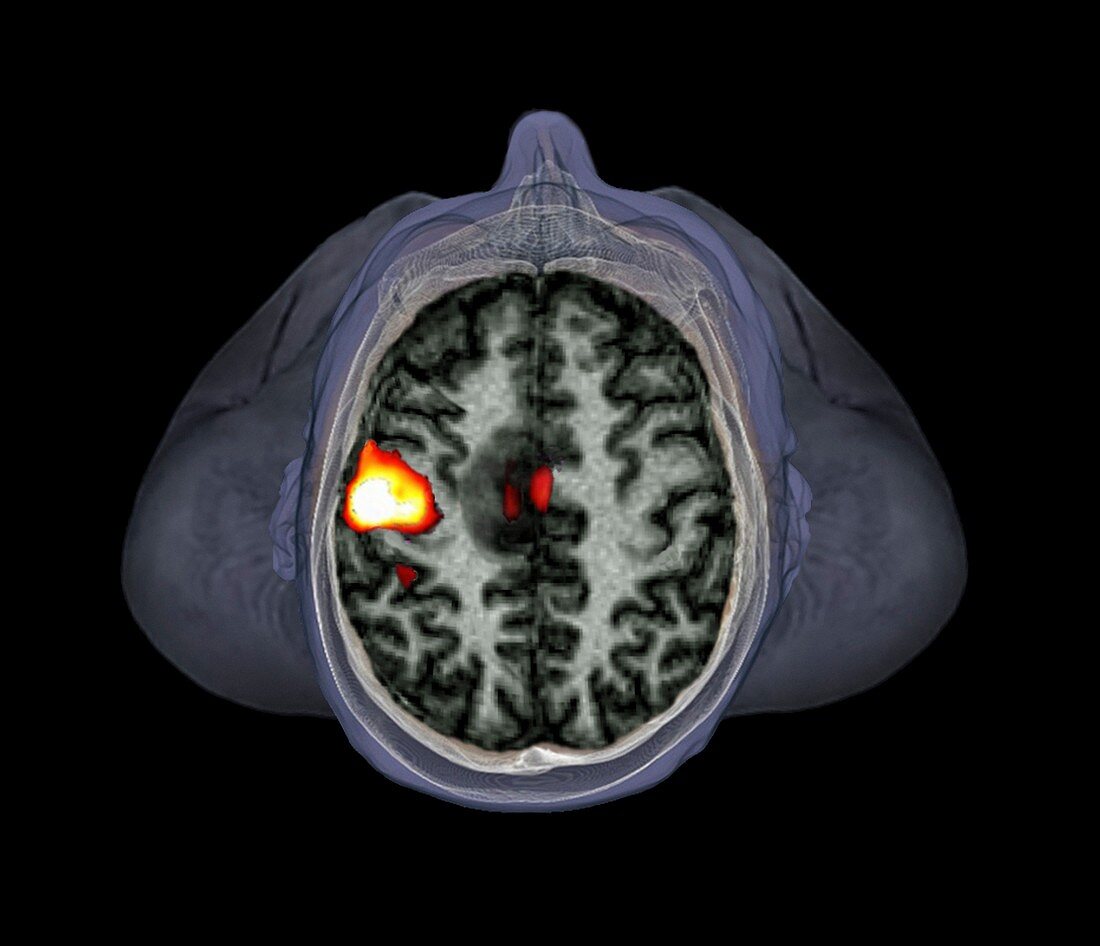
| Brain of a 39 year old man,functional magnetic resonance imaging (fMRI) scan (from above). This patient has a malignant (cancerous) tumour in the left hemisphere of the brain. FMRI is used to detect changes in blood flow and blood oxygenation in the brain,and to highlight areas of increased stimulation. Here,the yellow-orange area of the left hemisphere of the brain is part of the motor cortex,which is close to the lesion. It is showing increased activity due to the subject moving their right hand. This helps surgeons to identify the risks involved with surgical intervention | |
| Lizenzart: | Lizenzpflichtig |
| Credit: | Science Photo Library / Zephyr |
| Bildgröße: | 4518 px × 3886 px |
| Modell-Rechte: | nicht erforderlich |
| Eigentums-Rechte: | nicht erforderlich |
| Restrictions: | - |
Preise für dieses Bild ab 15 €
Universitäten & Organisationen
(Informationsmaterial Digital, Informationsmaterial Print, Lehrmaterial Digital etc.)
ab 15 €
Redaktionell
(Bücher, Bücher: Sach- und Fachliteratur, Digitale Medien (redaktionell) etc.)
ab 30 €
Werbung
(Anzeigen, Aussenwerbung, Digitale Medien, Fernsehwerbung, Karten, Werbemittel, Zeitschriften etc.)
ab 55 €
Handelsprodukte
(bedruckte Textilie, Kalender, Postkarte, Grußkarte, Verpackung etc.)
ab 75 €
Pauschalpreise
Rechtepakete für die unbeschränkte Bildnutzung in Print oder Online
ab 495 €
Keywords
- 30er Jahre,
- abnormal,
- Aktivität,
- Bewegung,
- Bildgebung,
- Diagnose,
- dreißiger Jahre,
- Erwachsene,
- farbig,
- FMRI,
- funktionellen Magnetresonanztomographie,
- gefärbt,
- Gehirn,
- Großhirn,
- Krebs,
- krebsartig,
- linke Hemisphäre,
- maligne,
- Malignom,
- Mann,
- Männlich,
- Medizin,
- medizinisch,
- Mensch,
- menschlicher Körper,
- MRT-Untersuchung,
- Neuroimaging,
- Neurologie,
- neurologisch,
- Onkologie,
- präoperativ,
- Schaden,
- schwarzer Hintergrund,
- Tumor,
- ungesund,
- Wunde,
- Zusammengesetzt