Blue mussel,light micrograph
Bildnummer 11681505
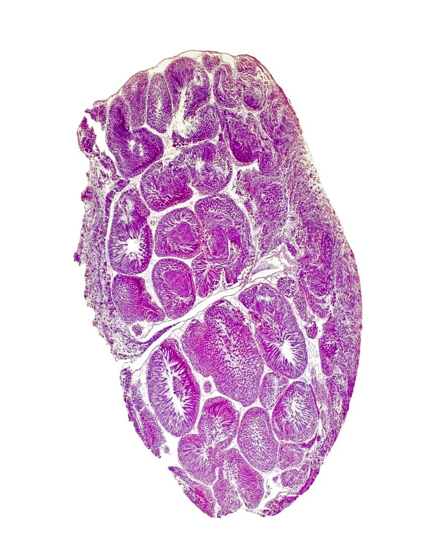
| Blue mussel. Light micrograph of a transverse section through the gonads and mantle of a male blue mussel (Mytilus edulis). The oval structures are seminiferous tubules,the site of spermatogenesis (sperm production). Mature sperm cells can be seen closest to each tubule's lumen (white),with their tails protruding into the lumen. Moving away from the lumen are increasingly immature precursor cells; first spermatids,then spermatocytes and finally spermatogonia. The outermost cells are sertoli cells,which nourish the maturing sperm. The mantle (left nad right) covers the soft parts of the mussel and lines the interior of the shell. It secretes calcium carbonate,which forms the shell. Magnification: x7 when printed at 10 centimetres tall | |
| Lizenzart: | Lizenzpflichtig |
| Credit: | Science Photo Library / Wheeler, Dr. Keith |
| Bildgröße: | 3553 px × 4441 px |
| Modell-Rechte: | nicht erforderlich |
| Eigentums-Rechte: | nicht erforderlich |
| Restrictions: | - |
Preise für dieses Bild ab 15 €
Universitäten & Organisationen
(Informationsmaterial Digital, Informationsmaterial Print, Lehrmaterial Digital etc.)
ab 15 €
Redaktionell
(Bücher, Bücher: Sach- und Fachliteratur, Digitale Medien (redaktionell) etc.)
ab 30 €
Werbung
(Anzeigen, Aussenwerbung, Digitale Medien, Fernsehwerbung, Karten, Werbemittel, Zeitschriften etc.)
ab 55 €
Handelsprodukte
(bedruckte Textilie, Kalender, Postkarte, Grußkarte, Verpackung etc.)
ab 75 €
Pauschalpreise
Rechtepakete für die unbeschränkte Bildnutzung in Print oder Online
ab 495 €