Surgery to fuse the cervical spine
Bildnummer 11681348
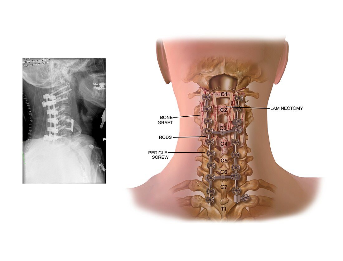
| Surgery to fuse the cervical spine. Labelled artwork of a posterior view of the cervical (neck) spine after surgery to fuse the spine and implant stabilizing rods. The X-ray at left shows a lateral (side) view of the rods after implantation. This operation is an example of internal fixation. In the artwork,the vertebrae are labelled (C1 to C7). Spinal rods have been implanted all the way from C1 to C7. The vertebrae from C1 to C4 have undergone a laminectomy,the removal of part of the lamina. The laminae are thin bony plates that form part of each each vertebra. Bone grafts (red) have also been used. The rods are held in place by pedicle screws | |
| Lizenzart: | Lizenzpflichtig |
| Credit: | Science Photo Library / Alesi, John T. |
| Bildgröße: | 4798 px × 3661 px |
| Modell-Rechte: | nicht erforderlich |
| Eigentums-Rechte: | nicht erforderlich |
| Restrictions: | - |
Preise für dieses Bild ab 15 €
Universitäten & Organisationen
(Informationsmaterial Digital, Informationsmaterial Print, Lehrmaterial Digital etc.)
ab 15 €
Redaktionell
(Bücher, Bücher: Sach- und Fachliteratur, Digitale Medien (redaktionell) etc.)
ab 30 €
Werbung
(Anzeigen, Aussenwerbung, Digitale Medien, Fernsehwerbung, Karten, Werbemittel, Zeitschriften etc.)
ab 55 €
Handelsprodukte
(bedruckte Textilie, Kalender, Postkarte, Grußkarte, Verpackung etc.)
ab 75 €
Pauschalpreise
Rechtepakete für die unbeschränkte Bildnutzung in Print oder Online
ab 495 €
Keywords
- abnormal,
- Arthrologie,
- ausgeschnitten,
- Ausschnitte,
- Behandlung,
- beschriftet,
- Betrieb,
- C1,
- C2,
- C3,
- C4,
- chirurgisch,
- Diagramm,
- Etikette,
- Etiketten,
- Gebärmutterhals-,
- geduldig,
- Gelenk,
- Gelenke,
- Hals,
- Illustration,
- Implantat,
- Joint,
- Knochen,
- Knochenspan,
- Kollabierende Wirbelsäule,
- Kondition,
- Kunstwerk,
- Laminektomie,
- Medizin,
- medizinisch,
- Mensch,
- Menschen,
- menschlicher Körper,
- Niemand,
- Operation,
- Person,
- posterior,
- Radiographie,
- Röntgen,
- Röntgengerät,
- Rückansicht,
- Rückgrat,
- Scheibe,
- Schrauben,
- Seitenansicht,
- seitlich,
- Stangen,
- Störung,
- Technologie,
- technologisch,
- Text,
- ungesund,
- vertebral,
- weißer Hintergrund,
- Wirbel,
- Wirbelsäule,
- Wirbelsäulen-