Fractured lumbar vertebral processes
Bildnummer 11681323
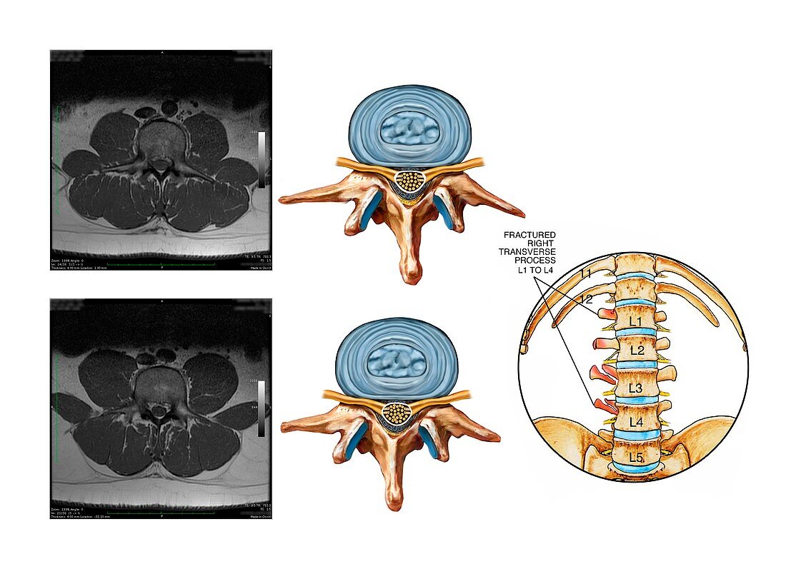
| Fractured lumbar vertebral processes. Labelled artworks and corresponding axial MRI (magnetic resonance imaging) scans of lumbar vertebrae (L3 and L4) with fractures of the transverse processes. The fractures (red) are shown in the circular inset at lower right. The artworks at centre (views from above) show the normal appearance of the vertebrae. This includes the spinal cord (yellow),the spinal nerves (yellow),the various bony projections of the vertebrae,and the discs of fibrocartilage (blue) that fill the space between each vertebra. The MRI scans at left also show the muscles (dark grey) of the spine and lower back that help protect the vertebrae | |
| Lizenzart: | Lizenzpflichtig |
| Credit: | Science Photo Library / Alesi, John T. |
| Bildgröße: | 4854 px × 3609 px |
| Modell-Rechte: | nicht erforderlich |
| Eigentums-Rechte: | nicht erforderlich |
| Restrictions: | - |
Preise für dieses Bild ab 15 €
Universitäten & Organisationen
(Informationsmaterial Digital, Informationsmaterial Print, Lehrmaterial Digital etc.)
ab 15 €
Redaktionell
(Bücher, Bücher: Sach- und Fachliteratur, Digitale Medien (redaktionell) etc.)
ab 30 €
Werbung
(Anzeigen, Aussenwerbung, Digitale Medien, Fernsehwerbung, Karten, Werbemittel, Zeitschriften etc.)
ab 55 €
Handelsprodukte
(bedruckte Textilie, Kalender, Postkarte, Grußkarte, Verpackung etc.)
ab 75 €
Pauschalpreise
Rechtepakete für die unbeschränkte Bildnutzung in Print oder Online
ab 495 €
Keywords
- abnormal,
- Anatomie,
- anatomisch,
- Arthrologie,
- ausgeschnitten,
- Ausschnitte,
- axial,
- Bandscheiben,
- beschriftet,
- Degeneration,
- Diagramm,
- Etikette,
- Etiketten,
- Fragmente,
- Fraktur,
- frakturiert,
- Gelenk,
- Illustration,
- Joint,
- kaputt,
- Knochen,
- Kondition,
- Kunstwerk,
- L1,
- L2,
- L3,
- L4,
- L5,
- links,
- Medizin,
- medizinisch,
- menschlicher Körper,
- Niemand,
- Prozess,
- Querschnitt,
- Recht,
- Rücken,
- Rückenmark,
- Rückgrat,
- Sektion,
- sektioniert,
- Störung,
- Text,
- ungesund,
- unterer Rücken,
- Verletzung,
- Von Oben,
- weißer Hintergrund,
- Wirbel,
- Wirbelsäule,
- Wirbelsäulen-