Slipped discs in the cervical spine
Bildnummer 11681297
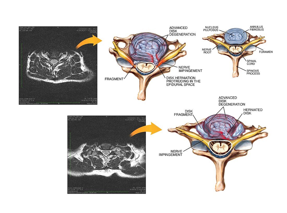
| Slipped discs in the cervical spine. Labelled artworks and corresponding axial MRI (magnetic resonance imaging) scans through the cervical (neck) spine showing slipped (herniated) discs at the C5-C6 joint (top) and the C6-C7 joint (bottom). A normal spinal joint is at top right. Fragments from the degenerating discs are pressing on cervical nerves (top) branching off the spinal cord or protruding into the C7 foramen (bottom). The front of the spine is at top in each view. The discs of fibrocartilage between each vertebra form joints that allow the spine to bend and flex. Spinal disc herniation can cause pain and in severe cases may require surgery | |
| Lizenzart: | Lizenzpflichtig |
| Credit: | Science Photo Library / Alesi, John T. |
| Bildgröße: | 4860 px × 3614 px |
| Modell-Rechte: | nicht erforderlich |
| Eigentums-Rechte: | nicht erforderlich |
| Restrictions: | - |
Preise für dieses Bild ab 15 €
Universitäten & Organisationen
(Informationsmaterial Digital, Informationsmaterial Print, Lehrmaterial Digital etc.)
ab 15 €
Redaktionell
(Bücher, Bücher: Sach- und Fachliteratur, Digitale Medien (redaktionell) etc.)
ab 30 €
Werbung
(Anzeigen, Aussenwerbung, Digitale Medien, Fernsehwerbung, Karten, Werbemittel, Zeitschriften etc.)
ab 55 €
Handelsprodukte
(bedruckte Textilie, Kalender, Postkarte, Grußkarte, Verpackung etc.)
ab 75 €
Pauschalpreise
Rechtepakete für die unbeschränkte Bildnutzung in Print oder Online
ab 495 €
Keywords
- abnormal,
- Anatomie,
- anatomisch,
- Arthrologie,
- ausgeschnitten,
- Ausschnitte,
- axial,
- Bandscheiben,
- Bandscheibenvorfall,
- beschriftet,
- C5,
- C6,
- C7,
- Diagnose,
- Diagramm,
- Etikette,
- Etiketten,
- Fragment,
- Fragmente,
- Gebärmutterhals-,
- Gelenk,
- Gelenke,
- gesund,
- Hals,
- Illustration,
- Joint,
- Knochen,
- Kondition,
- Kunstwerk,
- Magnetresonanztomografie,
- Medizin,
- medizinisch,
- menschlicher Körper,
- MRT-Untersuchung,
- Nerven,
- Nervenkompression,
- Nervensystem,
- neural,
- Niemand,
- normal,
- Querschnitt,
- Rückenmark,
- Rückgrat,
- Schmerz,
- schmerzhaft,
- Sektion,
- sektioniert,
- Störung,
- Text,
- ungesund,
- Vergleich,
- vergleichen,
- Verletzung,
- vertebral,
- weißer Hintergrund,
- Wirbel,
- Wirbelsäule,
- Wirbelsäulen-