White blood cell,X-ray diffraction image
Bildnummer 11680570
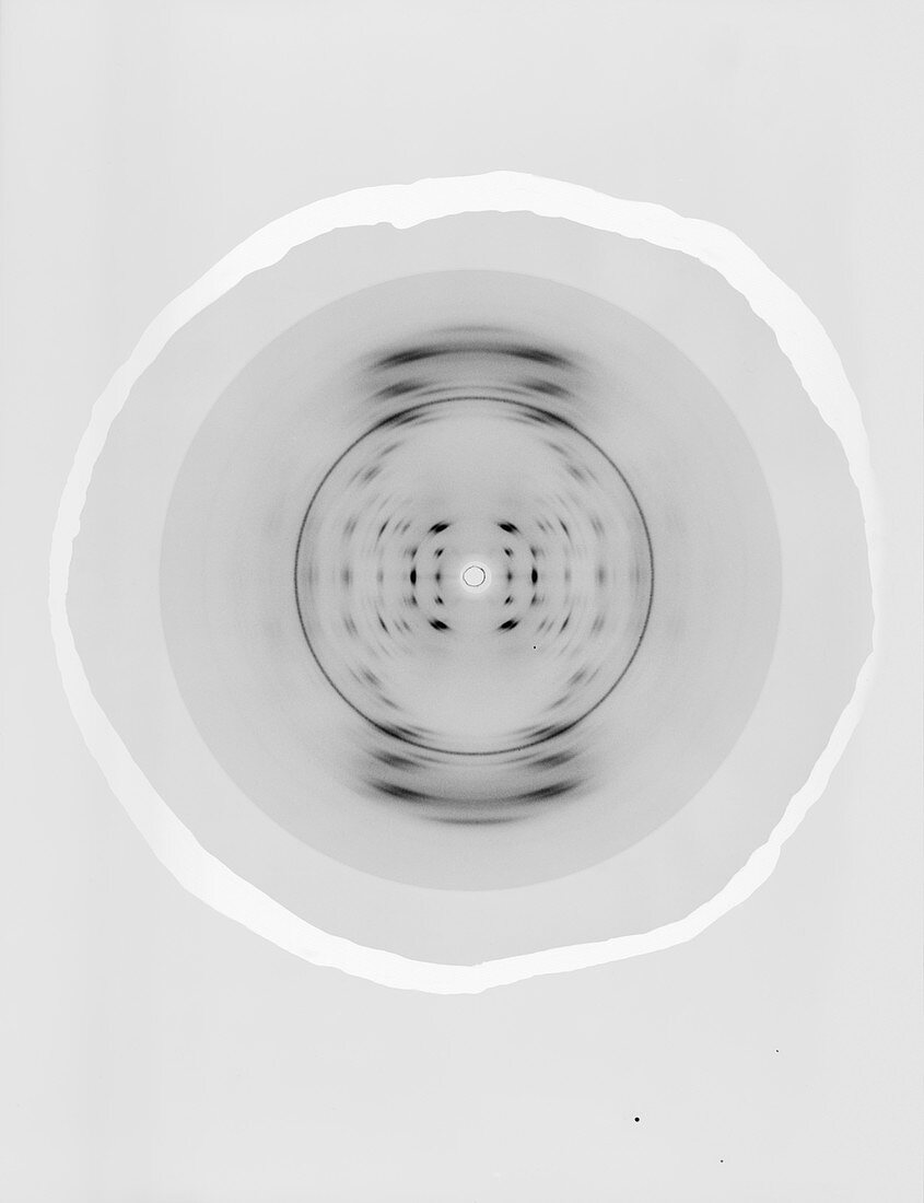
| White blood cell. X-ray crystallography diffraction pattern for a white blood cell,obtained as part of research on biochemical structures. This image was produced by British physicist and molecular biologist Maurice Wilkins (1916-2004) while working at the MRC Biophysics Research Unit at King's College London,UK. It was here in the early 1950s that Wilkins,Rosalind Franklin,and other crystallographers obtained X-ray diffraction patterns for DNA that led to James Watson and Francis Crick's DNA double helix model. As well as DNA,the King's College researchers investigated other materials,cells,and biological tissues | |
| Lizenzart: | Lizenzpflichtig |
| Credit: | Science Photo Library / King's College London |
| Bildgröße: | 3697 px × 4813 px |
| Modell-Rechte: | nicht erforderlich |
| Eigentums-Rechte: | nicht erforderlich |
| Restrictions: | - |
Preise für dieses Bild ab 15 €
Universitäten & Organisationen
(Informationsmaterial Digital, Informationsmaterial Print, Lehrmaterial Digital etc.)
ab 15 €
Redaktionell
(Bücher, Bücher: Sach- und Fachliteratur, Digitale Medien (redaktionell) etc.)
ab 30 €
Werbung
(Anzeigen, Aussenwerbung, Digitale Medien, Fernsehwerbung, Karten, Werbemittel, Zeitschriften etc.)
ab 55 €
Handelsprodukte
(bedruckte Textilie, Kalender, Postkarte, Grußkarte, Verpackung etc.)
ab 75 €
Pauschalpreise
Rechtepakete für die unbeschränkte Bildnutzung in Print oder Online
ab 495 €
Keywords
- 1900er Jahre,
- 20. Jahrhundert,
- Analyse,
- analytisch,
- Biochemie,
- biochemisch,
- Biologie,
- biologisch,
- Einfarbig,
- Geschichte,
- historisch,
- Kristallographie,
- Maurice Wilkins,
- menschlicher Körper,
- Molekül,
- Molekularbiologie,
- Niemand,
- Physik,
- physikalische Chemie,
- physisch,
- Röntgen,
- Schwarz und weiß,
- Struktur,
- strukturell,
- weißes Blutkörperchen,
- Zelle